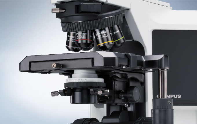
BX43 현미경은 고성능 광학계와 디지털 이미징, 폭넓게 다양한 특징을 제공합니다. 이 유연한 현미경은 뛰어난 연색성(演色性)을 위한 트루컬러 LED 조명과 결합한 다양한 관찰 방법과 뛰어난 광학계를 제공합니다. 각 대물렌즈에 맞추어 광량을 조절해야 하는 일을 없애주는 Light Intesity Manager에 의한 최적화된 쉬운 사용법으로 그 효율성이 극대화됩니다. 넓은 범위의 액세서리들은 그 시스템을 더 많은 기술을 커버할 수 있도록 확장해주고, 미래의 요구들을 만족하게 하도록 맞춰줍니다.
사용자를 위한 인체 공학
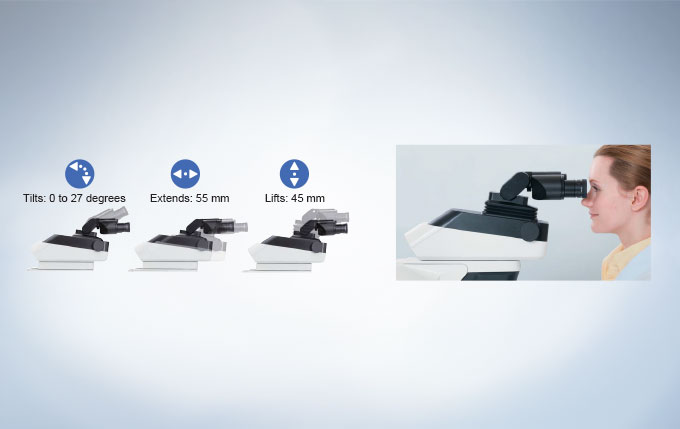
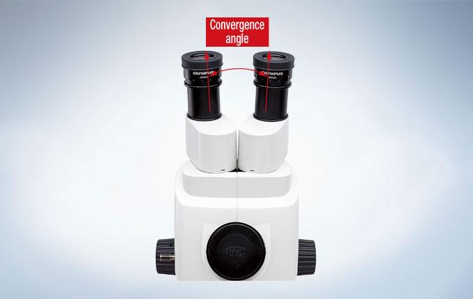
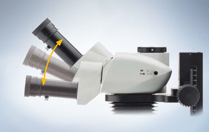
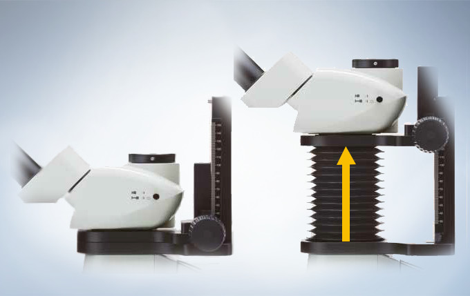
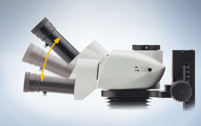
안정된 관측 자세는 사람마다 다르므로, 이 현미경은 각 사용자에 맞춰 mm 단위의 조절이 가능합니다. Olympus 경통 제품군은 경사각, 경통 연장 혹은 높이 조절과 같은 기능을 제공하여 완벽한 3차원적으로 유연한 조절이 가능합니다.
높은 조절성의 이안 경통
현미경 사용 시, 바르면서 편안한 자세를 취하는 것은 필수적이나, 전적으로 사용자에게 달려있는 부분입니다. 인체 공학적으로 완벽한 거리, 꺾임, 높이 조절이 가능한 경통은 경사각, 길이와 높이 조절이 가능, 3차원적으로 완벽하게 유연한 설정이 가능합니다. 그 결과,다른 방법 보다 정확하게 현미경을 사용자에 맞도록 조절할 수 있게 되었습니다.
모든 요구를 위한 이안 경통
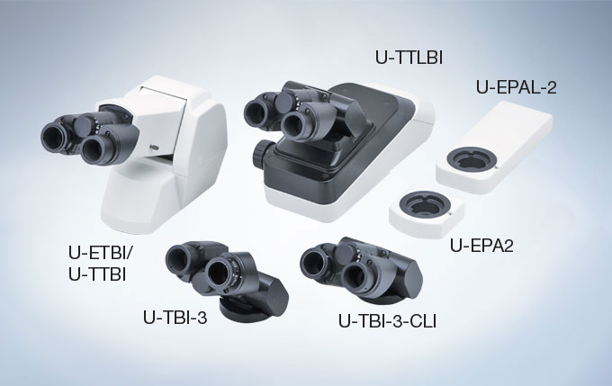
다양한 이안 경통 제품 군은 다양한 요구를 만족시킬 수 있습니다. 어떤 모델은 일반적인 도립상을 생성하는 반면 다른 모델은 표본과 동일한 방향으로 움직이는 정립상을 보여주어 표본 상에서 특정 부위를 보다 쉽게 찾을 수 있습니다.
효율성 증가를 위한 디자인
균일한 밝기를 위한 Light Intensity Manager
시야 범위에서의 전반적인 밝기 유지를 위해서는 각 배율에 맞는 조명의 세기가 필요하다는 것은 광학적 사실입니다. 시료의 스크리닝(Screening) 작업 시, 대물렌즈 배율을 아래위로 반복하여 움직이는 것은 즉, 대물렌즈 변환 장치를 돌릴 때마다 조명의 광량을 조절해주어야 한다는 것은 일반적인 일입니다. 천연색 LED와 함께 사용되는 Light Intensity Manager는 각 대물렌즈 변경에 맞추어 사용자가 미리 설정한 수준으로 조명 광량을 자동 조절합니다.
병리 및 세포학을 위한 최적의 광원
Olympus는는 진보된 혼합기질 LED 기술로 차세대 명시야(Bright Field) 조명을 이끌고 있습니다. 이 천연색 LED는 그 색상 표현 지표가 주광색 필터(Daylight filter)를 장착한 할로겐 램프와 유사하도록 디자인 되어져 왔습니다. 이들이 생성하는 색상은 천연색 LED와 주광색 필터(Daylight filter) 하의 할로겐 램프가 정확히 일치하는 모습을 띄고 있습니다. 게다가 비슷한 색상들이 명확하게 구별될 수 있습니다. 이러한 진보된 색상 표현 기술은 주로 사용되는 대부분의 염색 기법에 이상적 파장대역을 제공합니다. (예시. haematoxylin/eosin – HE와 Papanicolaou – Pap)
저 배율 집광기 U-LC
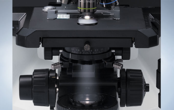
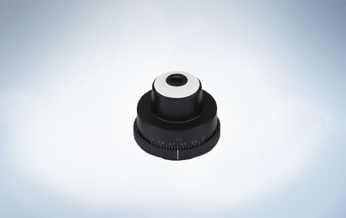
넓은 범위 집광기 U-LC는 임상 분야에서의 명시야(Brightfield) 검경에 적합하도록 설계되었습니다. 이들의 광학 설계는 2x ~ 100x 사이의 배율 범위 사용이 가능하게 합니다. 이는 대물렌즈 변경 시 광학계 밖으로 상단 렌즈(Top lens)를 넣었다 뺐다 할 필요가 없도록 하여 사용자를 더욱 효율적이고 편안하게 만들어 줍니다.
많은 어플리케이션들을 위한 확장성
BX43은 임상 시장을 위한 뛰어난 다양한 특징과 광학 성능을 제공합니다. 강력한 UIS2 광학계 시스템, 탁월한 강도의 쉬운 조작성의 전면부 제어 방식을 채용한 탁월한 강도의 Y자 본체 그리고 독특한 색재현성의 LED 명시야(Bright Field) 조명은 다재다능하고 인체 공학적 조작을 가능하게 합니다. 효율적인 공간활용도의 BX43 현미경 본체는 뛰어난 가성비를 바탕으로 쉬운 반복 작업이 가능하고, 다양한 용도로 사용되는 stand-alone 현미경으로서 기본적인 영상작업에 적합합니다 – 진보된 요구를 만족시키기 위해 준비된 성능과 다재다능함.
시스템의 융통성
BX43은 특화된 전문가와 유연한 시스템 사이에서 이상적 균형을 유지하는 임상 스크리닝(Clinical Screening) 환경을 제공합니다. 긴 시간 동안의 명시야(Bright Field) 스크리닝(Screening) 작업에 적합하며 BX46과 동일한 UIS2 광학계와 향상된 색 재현성의 LED 기술은 최고의 선명도를 제공합니다.
모든 어플리케이션들을 위한 집광기
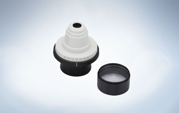
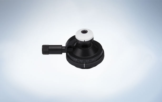
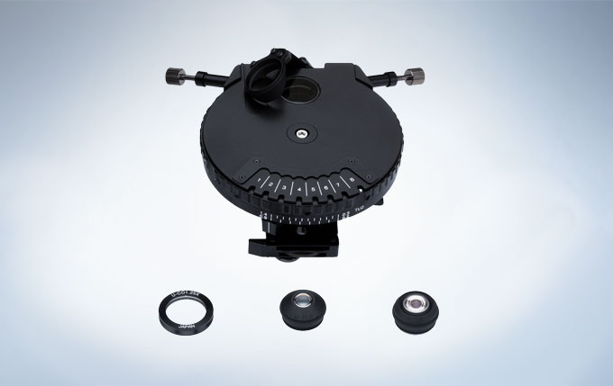
Olympus는 수 많은 어플리케이션에 적합하도록 UIS2 집광기 제품군을 개발해왔습니다. 염색된 시료의 명시야(Bright Field) 관찰 (HE 염색 조직 절편과 같은)이 핵심인 임상 분야를 위해 Olympus는 육안으로도 쉽게 슬라이드를 배치가 가능한 흰색 표면의 집광기를 개발해왔습니다. BX43은 명시야(Brightfield_ BF), 암시야(Darkfield_DF) 그리고 위상차(Phase Contrast_PH) 관찰을 지원함과 동시에 저 배율을 위해 특별히 설계된 집광기를 장착할 수 있습니다. 추가적으로 유니버셜 집광기는 암시야(Darkfield)와 위상차(Phase Contrast) 그리고 1.25X 배율을 위한 특수 삽입 장치가 포함되어 이 모든 방법들이 가능합니다. 넓은 범위의 집광기는 임상에서의 명시야(Bright Field) 관측에 필요한 요구사항을 만족시키기 위해 설계되어왔습니다.
교육과 토론에 이상적
새로운 인원의 합류 시, 가장 중요한 점은 기존 팀원으로 부터 배울 수 있어야 한다는 것입니다. 이를 가능한 효율적으로 만드는 것은 모든 교육 시설의 핵심 목표입니다. 실제 상황에서 팀 토론과 사례 회의는 매우 중요하며 이를 촉진하는 일은 특별한 다른 해결책들이 필요합니다.
그룹 관측 시스템
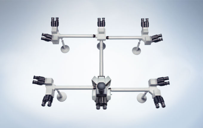
인체 공학적인 이안 경통, 삼안 경통 뿐만 아니라 Olympus는 사용자에 맞게 최적화할 수 있는 실험실 토론용 이인, 다중 관측 연결 장치를 공급 합니다. 이 시스템은 모든 그룹원들이 각자 접안렌즈로 동시에 표본을 보고 토론할 수 있으므로 임상·교육적 관찰에 적합합니다. 2~10명 혹은 그 이상의 검경을 위해 폭넓게 선택할 수 있도록 설계되었습니다.
다양한 디지털 이미징 액세서리
다재다능한 BX43 시스템은 어떠한 어플리케이션에도 맞게 변형될 수 있는 잠재력을 가진 현미경입니다. 최첨단 연구부터 컨퍼런스에 최적화된 독립형(Stand-alone) 모델까지 당사의 디지털카메라와 cellSens 이미징 소프트웨어의 완벽한 제품군은 모든 임상학적 진단 시 정교한 픽셀의 색상 표현력으로 조직 염색 영상 획득을 보장합니다.
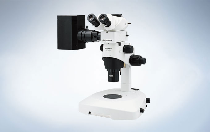
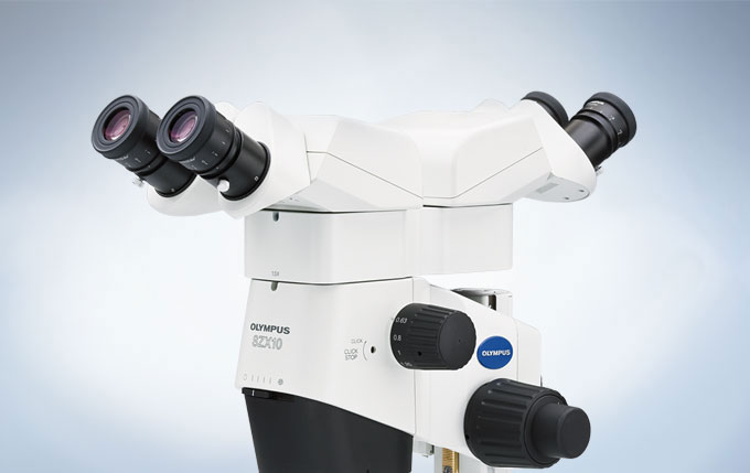
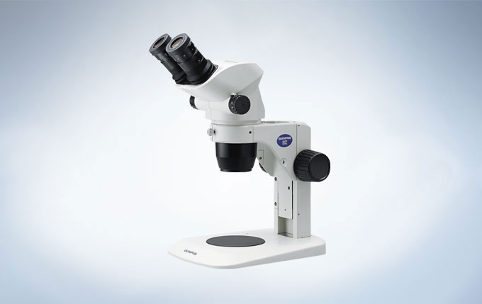
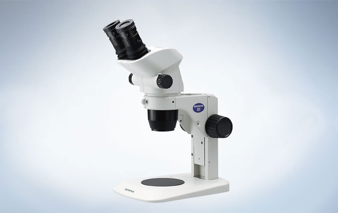
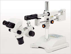
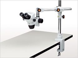
SZX10 은 특히 일상적인 연구를 위해 설계되었습니다. 0.2의 최대 개구수(NA, Numerical Aperture)로 1mm 내 600줄을 구별하는 해상력과 10:1의 줌 비율은 표본으로부터의 더 많은 정보의 효율적 수집이 가능하게합니다. 뛰어난 작동거리(Working Distance)를 제공하는 대물렌즈의 넓은 선택의 폭과 갈릴리언(Galilean) 광학 시스템은 자연스럽고 왜곡 없는 관찰이 가능합니다. 이 혁신적인 디자인은 시야의 편안함과 인체 공학적 사용을 보장합니다. SZX10는 오블리크(Oblique), 명시야(Brightfield) 조명에 적합하고 여러 이미징 장치와의 호환성은 형광 및 유연한 이미징 스테이션으로서 활용을 가능하게 합니다.
넓은 줌 배율의 다용도 실체 현미경
우수한 광학 성능과 자연 그대로의 관찰
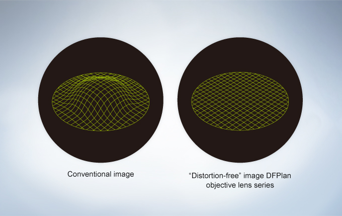
SZX10 은 뛰어난 입체감, 색상 표현으로 표본의 원래 모습을 완벽하게 재현하도록 설계되었습니다. DFPlan 시리즈는 높은 NA 및 해상력과 사용의 편이성과 평탄도 높은 이미징을 위한 “왜곡 없는” 광학계 디자인을 결합되어 2x 배율 대물렌즈(NA 0.2)로 1mm 내 600줄을 구별하는 고해상도 이미징이 가능합니다.
10:1의 광폭 줌 배율
SZX10은 1x 대물렌즈로 6.3x ~ 63x 배율 범위를, 2x 대물렌즈로 123x 배율까지 대응합니다. 이 10:1의 광폭 줌 배율은 해부 및 표본 조작을 위한 저배율 접사(Macro) 관측부터 세포 구조의 선명한 고배율 관측까지의 모든 생명 과학 어플리케이션에 적합합니다.
단순 형광 관측
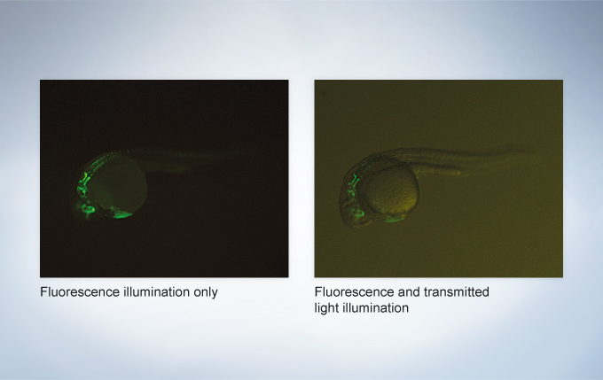
형광 유닛을 추가하여 SZX10으로 생체 내에서 발현된 형광 단백질을 관찰할 수 있습니다. 이 유닛의 고성능 형광 필터 구성품은 빠른 시작과 영상 품질 향상을 위한 높은 형광 검출 효율을 제공합니다.
디지털 이미징과 문서화
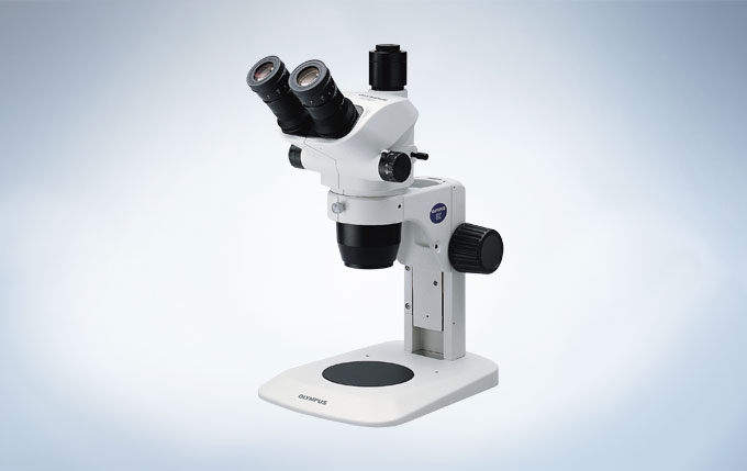
SZX10은 삼안경통, Olympus DP 시리즈 디지털카메라와 함께 사용하여 고해상도 이미징과 연구용 문서작업이 가능합니다. DP 시리즈 카메라는 높은 감도가 요구되는 형광 이미징을 포함, 다양한 어플리케이션을 고감도로 관측하고, Olympus cellSens 소프트웨어로 손쉽게 제어할 수 있습니다.
일상적인 작업을 위한 인체 공학적 디자인
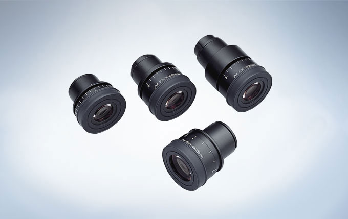
눈의 피로를 줄인 ComfortView 접안렌즈
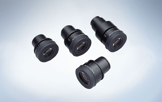
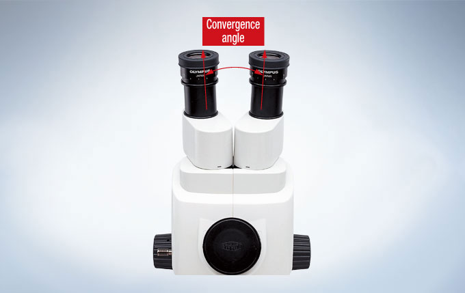
ComfortView 접안렌즈로 3D 이미지 관측 시, 안구 운동 범위가 넓어 단기 및 장기 사용이 매우 편안합니다. 또한 이것은 입체 영상의 형성을 쉽게 하여, 눈의 피로를 덜어주는 결과를 가져옵니다. 더불어, 뛰어난 광학 코팅은 세부사항까지 정확한 색으로 표현합니다.
인체 공학적 경통
기울기 조정이 가능한 꺽임형 이안경통(5 ~ 45도 기울기)은 사용자에게 가장 편안한 눈 위치를 신속하게 찾아내고 척추와 척추기립근의 부담을 최소화 하여 생산성과 일상적 어플리케이션의 품질을 향상시킵니다.
울트라 슬림 LED 베이스(Ultra-Slim LED Base)와 오블리크(Oblique) 조명
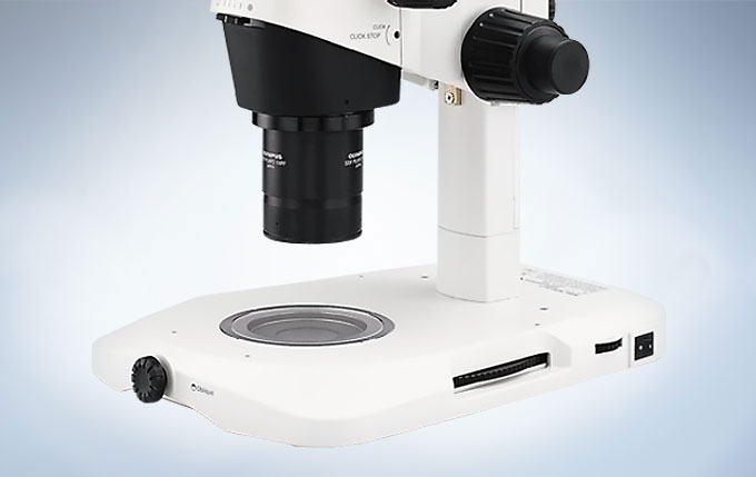
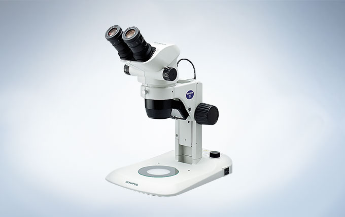
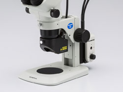
LED 베이스 시스템은 밝고 고른 영상을 위해 투과 조명의 미세 조절을 제공합니다. 울트라 슬림 사이즈의 40mm 두께 베이스는 인체공학적으로 뛰어난 재물대 높이를 제공합니다. 새로운 원형 컨베이어 교환 장치는 명시야(Brightfield)와 암시야(Darkfield)의 조명 방식의 선택과 제어를 편리하게 만들어 주고, 긴 수명의 LED는 재물대와 표본에 열을 주지 않습니다. LED 조명 시스템은 매우 정확하고 완벽하게 고른 오블리크(Oblique) 조명을 만들기 위해 얇은 판 구조의 미세 셔터 시스템을 이용합니다.
모든 용도를 위한 액세서리
고성능 대물렌즈의 선택
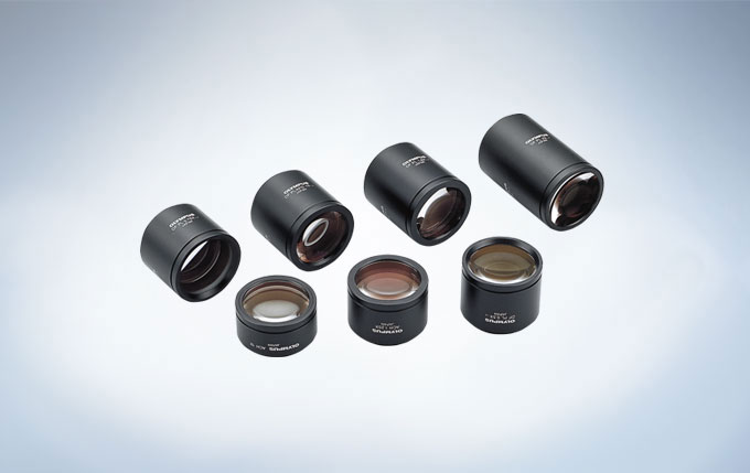
DFPL2x/ DPFL1.5x/ DFPL 0.75x/ DFPL 0.5x DFPL 시리즈 대물렌즈는 표본의 색과 형태를 정확하게 재현합니다. DFPLAPO 1.25x /DFPLAP01x4 최고 등급의 성능을 얻기 위한 Apochromat 보정 뛰어난 해상력과 대비, 그리고 왜곡이 최소화된 영상의 평탄도. SZX-ACH1.25x /SZX-ACH1x 높은 해상력과 긴 작동거리(Working distance)의 균형 잡힌 대물렌즈의 제공.
최적의 조명 베이스 선택
SZX2-ILLT 일반 명시야(Brightfield)와 대비 강조 명시야(Brightfield), 암시야(Darkfield) 그리고 오블리크(Oblique) 조명들을 위한 울트라 슬림 투과광 솔루션 밝은 LED 베이스는 일반 베이스의 절반 높이인 41 mm이고, 놀라운 유연성 및 숙련자와 초보자 모두에게 적합한 인체 공학적 형태와 견고함을 제공합니다.
SZX2-ILLK 명시야(Brightfield)와 오블리크(Oblique) 조명을 위한 투과광 스탠드 6 V/ 30 W 할로겐 램프가 내장되어 투명한 표본의 Contrast가 향상된 이미지를 제공합니다.
SZX2-ILLD 명시야(Brightfield)와 암시야(Darkfield) 조명을 위한 투과광 스탠드 6 V/ 30 W 할로겐 램프가 내장되어 있고 명시야(Brightfield)와 암시야(Darkfield) 조명 전환이 가능하여, 배경 노이즈의 감소 및 대비의 개선으로 보다 섬세하게 세부사항을 볼 수 있습니다.
SZX2-ILLB 명시야(Brightfield) 및 오블리크(Oblique) 조명을 위한 고성능 투과광 스탠드 고/저대비의 선택은 선명하고 효율적인 대비와 조명 능력을 제공합니다. 빛 강도 및 색온도는 6 V/ 30 W 할로겐 램프 상에서 쉽게 조절될 수 있습니다. 고배율 대물렌즈를 사용하여 대비가 강조된 구조들의 아주 미세한 부분까지 관찰 가능합니다. 특히 이쁜꼬마선충(C. elegans), 난모세포(Oocytes), 배아(Embryos) 관찰에 효과적입니다.
토론용 경통(Discussion Tube)
대면, 토론-스타일 중간 경통은 주관찰자와 보조관찰자가 서로 마주 앉은 상태에서 표본을 관측할 수 있게 해줍니다. 보조관찰자가 주관찰자의 작업을 보다 효과적으로 도울 수 있습니다.
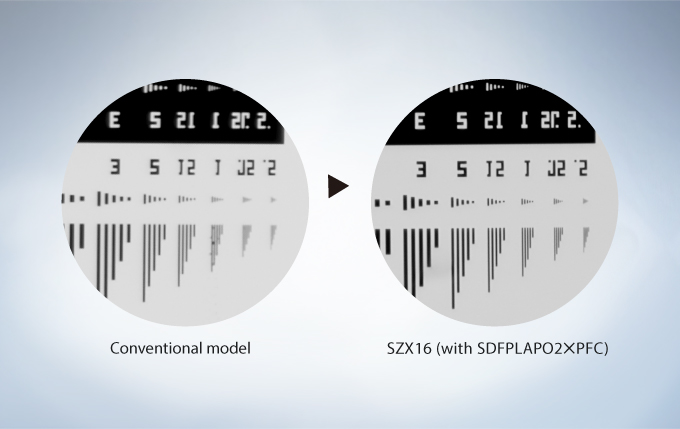
SZX16는 높은 수준의 연구용으로 디자인되었으며, 오블리크(Oblique), 명시야(Brightfield) 조명에 적합합니다. 0.3의 최대 개구수(NA, Numerical Aperture)는 1mm 내 900줄을 구분하는 해상력을 지원하므로 보다 많은 표본의 정보를 효율적으로 얻을 수 있습니다. 줌 비율이 16.4:1로 넓고, 우수한 작동거리(Working Distance)의 동초점(Parfocal) 대물렌즈를 사용하기 때문에 접사(Macro)와 마이크로(Micro) 관측의 전환이 쉽습니다. 자연스러움과 왜곡이 없는 시야를 제공하는 광학계와 함께 전체 개체 및 개별 세포 구조의 이미징은 쉬운 작업이 됩니다.
연구 실체현미경 시스템
울트라 와이드 줌과 함께하는 뛰어난 입체감
뛰어난 광학계 성능
경통과 Zoom Body 그리고 대물렌즈를 포함한 SZX16 광학계는 수차를 보정하여 모든 배율 범위에서의 수차로 인한 흐림(Chromatic blur) 현상을 보정하였습니다. SDF(Super Depth of Focus) 대물렌즈는 거의 모든 표본으로부터 선명하고 높은 대비의 영상을 가져올 수 있습니다. 최대 0.3의 개구수(NA)로 밀리미터 당 900줄을 구분할 수 있는 해상도는 아주 작은 세포와 세포 소기관의 구조에 대한 선명한 시각화가 가능합니다. 이처럼 놀라운 해상도와 뛰어난 배율은 작업을 효율적이고 정확하게 하여 표본으로부터 더욱 많은 정보를 얻게 해줍니다.
16.4:1 울트라 와이드 줌
SZX16의 울트라 와이드 줌 16.4:1은 1x 대물렌즈로 7x ~ 115x 배율을, 2x 대물렌즈로 230x 배율까지 가능합니다. 이 뛰어난 Zoom 범위로 해부 및 표본 조작을 위한 저배율 접사(Macro)부터 미세구조의 근접관측을 위한 섬세한 고배율 관측과 같은 다양한 연구 어플리케이션이 가능합니다.
3.5x ~ 230x 줌 배율을 위한 두 개의 대물렌즈
동초점 대물렌즈와 2구 회전 노스피스(Nosepiece)와 함께, 대물렌즈 교환이 매우 빠르고 쉬우며, 약간의 초점 재조정만 필요합니다. 그러므로 0.5x 및 2.0x 대물렌즈의 간섭 없는 배율 범위는 3.5x ~ 230x 배율입니다. 이는 효과적인 줌 비율 65.7:1을 대변합니다.
뛰어난 형광 관측
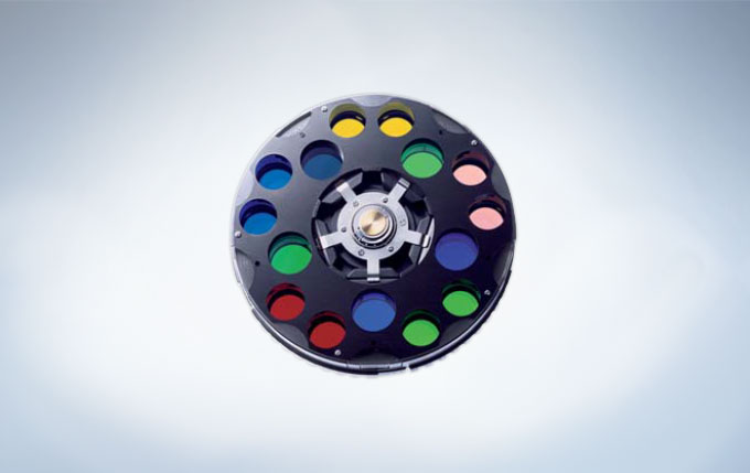
SZX16은 높은 개구수(NA)의 정확하게 설계된 광학계로 인해 한 개체 전부의 모습뿐만 아니라 핵 세부 구조까지 관측 가능한 형광 현미경으로 이상적입니다. 낮은 자가형광(Autofluorescence) 및 새 표면 코팅 기술을 적용한 고급 유리 소재로 인해 투과율 및 영상의 선명도가 크게 향상되었습니다. 이러한 특징들은 뛰어난 신호대잡음비와 높은 형광 신호 강도라는 결과를 가져옵니다. 저배율, 고해상도 이미징을 위하여 유연한 조명이 가능한 5 구 필터 휠에 의해 여기광(Excitation light)의 완벽한 조절이 편리해집니다.
모든 어플리케이션에서의 고해상력
두꺼운 초점 심도와 함께하는 고해상력
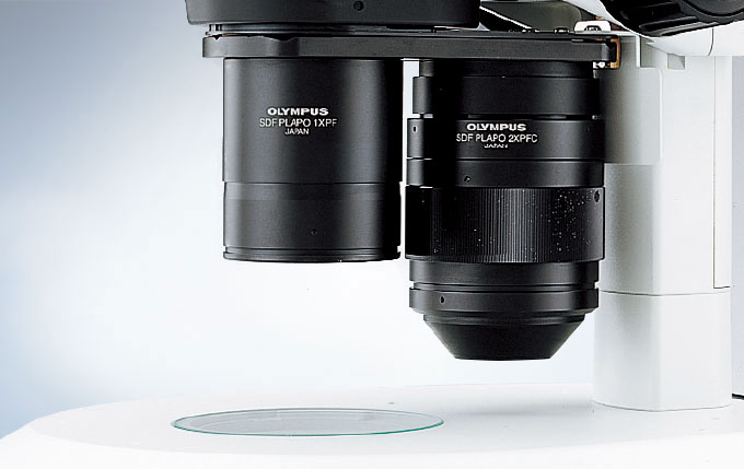
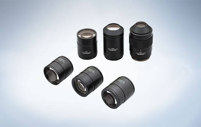
SDF(Super Depth of Focus) 대물렌즈는 거의 모든 표본으로부터 선명하고 높은 대비의 영상을 가져올 수 있습니다. SZX16은 6종류의 SDF 대물렌즈를 사용할 수 있으며, 이중 4종류는 동초점(Parfocal) Plan apochromat 0.5x, 1.0x, 1.6x, 2.0x 입니다) 0.3x 형석(Fluorite) 및 0.8x plan apochromat 대물렌즈는 보다 긴 작동거리(Working Distance)를 제공합니다.
배지 내 표본 관측에 최적화
SZX16용 2.0x SDF 대물렌즈는 0.3이라는 최고의 개구수(NA)를 가지고 있습니다. Plan apochromat 대물렌즈는 배양된 조직과 세포 관찰에 적합합니다. 이 Correction Ring은 용기와 매질의 굴절률 차이에 의해 발생하는 부조화를 미세하게 교정할 수 있습니다.. 이것은 페트리 디쉬 및 기타 배양 용기를 사용할 경우에도 최대 해상도에 이르는 것이 가능하도록 도와줍니다.
뛰어난 형광 성능
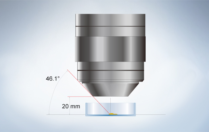
SDF 대물렌즈의 뛰어난 개구수(NA)특성은 형광 감도를 비약적으로 개선시켜줍니다. 또한, 새롭게 디자인된 수직에 가까운 반사 조명의 여기광 경로는 관찰 경로와 독립되어 있어 여기광의 효율성이 개선시킵니다. 이러한 기능 덕분에 모든 배율에서 기존 실체 현미경보다 더욱 밝은 형광 관찰이 가능합니다. 표본 검증을 위한 투과광 관측이 반사광 형광 관측 중에도 가능합니다.
SZX16 전동 초점 드라이브는 핸드 스위치를 통해 초점과 확대 모두를 간편하게 제어합니다. 이를 이용하여 EFI(Extended Focal Imaging)를 포함 디지털 문서화를 효율적이고 자동화할 수 있습니다. 이는 모의 3D 이미지 제작이 가능하며, 문서와 실제 눈으로 관측한 영상의 격차를 줄입니다. 자동 검사를 쉽게 하도록, Olympus cellSens 소프트웨어는 간단한 2D 측정부터 복잡한 위상 분석들을 사용하기 쉬운 환경에서 분석할 수 있도록 하기 위한 모든 도구를 제공합니다.
조작을 위한 충분한 작업 공간
현미경 상에서의 어떤 조작이든지 간에 표본 상단과 대물렌즈 하단 사이의 충분한 공간이 요구됩니다. SZX16에 사용 가능한 대물렌즈는 작동거리(Working Distance)가 길어서 수동 및 자동 조작과 주입 도구 모두를 사용할 수 있습니다. 그리고 1.6x, 2x와 같은 고배율 대물렌즈의 선단은 가늘어지기 때문에, Manipulator의 이동 범위는 눈에 띄게 넓어집니다.
눈의 피로를 줄인 ComfortView 접안렌즈
ComfortView 접안렌즈로 3D 이미지 관측 시, 안구 운동 범위가 넓어 단기 및 장기 사용이 매우 편안합니다. 이는 입체 이미지 형성을 보다 용이하게 하며, 결과적으로 눈의 피로를 덜어줍니다. 또한, 뛰어난 광학 코팅은 세부사항까지 정확한 색으로 표현합니다.
인체 공학적 경통
기울기 조정이 가능한 꺽임형 이안경통(5 ~ 45도 기울기)은 사용자에게 가장 편안한 눈 위치를 신속하게 찾아내고 등 및 근육의 부담을 최소화 하여 생산성과 일상적인 어플리케이션의 품질을 향상시킵니다.
액세서리의 다양함
최적 조명 베이스의 선택
SZX2-ILLT 일반 명시야(Brightfield)를 위한 울트라 슬림 투과광 솔루션으로 명시야(Brightfield), 암시야(Darkfield)와 오블리크(Oblique) 조명으로 대조를 향상하였습니다. 밝은 LED 베이스는 일반 베이스보다 절반 높이인 41 mm이고, 숙련된 사용자와 초보자에게 적합한 놀라운 유연성, 인체 공학 및 견고함을 제공합니다.
SZX2-ILLK 명시야(Brightfield)와 오블리크(Oblique) 조명을 위한 투과광 스탠드 입니다. 6 V/ 30 W 할로겐 램프가 내장되어 투명한 표본의 대조가 향상된 이미지를 제공합니다.
SZX2-ILLD 명시야(Brightfield)와 암시야(Darkfield) 조명을 위한 투과광 스탠드 입니다. 6 V/ 30 W 할로겐 램프가 내장되어 있고 명시야(Brightfield)와 암시야(Darkfield) 조명 전환이 가능하여, 배경 노이즈를 감소 및 대비를 개선하여 보다 섬세하게 세부사항을 볼 수 있습니다.
SZX2-ILLB 명시야(Brightfield) 및 오블리크(Oblique) 조명을 위한 고투과율 스탠드. 고/저대비의 선택은 선명하고 효율적인 대비와 조명 능력을 제공합니다. 6 V/ 30 W 할로겐 램프의 빛 강도 및 색온도 조절이 용이합니다. 고배율 대물렌즈를 사용하여 대비가 강조된 구조들의 아주 미세한 부분까지 관찰 가능합니다. 특히 이쁜꼬마선충(C. elegans), 난모세포(Oocytes), 배아(Embryos) 관찰에 효과적입니다.
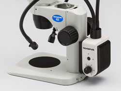
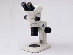
SZX7의 갈릴리언(Galilean) 광학 시스템은 동급에 비해 뛰어나며 디지털 이미징에 대해 동급 모델 대비 우수합니다. SZX7은 개선된 무한 보정 광학계와 7:1의 넓은 줌배율(0.8x~5.6x) 덕분에 1mm 내 최대 600줄을 구분하는 고해상도 영상을 높은 색 재현성(Color Fidelity)과 함께 제공합니다. 대물렌즈를 폭 넓게 선택할 수 있는 이 유연한 모듈식 디자인은 뛰어난 작동거리(Working distance)를 보증합니다. 새로운 컴포트뷰 접안렌즈를 통해 빠르고 편안한 검경이 보장되고, 인체공학적 디자인은 일상적인 어플리케이션들에서의 생산성과 품질을 향상시킵니다. 범용 LED 조명 스탠드는 LED 기술의 모든 이점을 함께 가져옵니다.
시스템 모듈 방식의 경제적인 스테레오 현미경
갈릴리언(Galilean) 광학 시스템
갈릴리언(Galilean) 두 개의 독립되고 평행한 줌 광학계를 채용하여 광학 성능과 시스템 모듈 유연성이 뛰어납니다.신중하게 선택된 렌즈 표면 코팅과 Apochromatic Zoom 광학계는 표본을 실제 색상과 최대한 유사한 컬러로 관측하고 문서화할 수 있습니다.
7:1 와이드 줌
1x 대물렌즈로 Zoom 배율 8x ~ 56x 배율을, 2x 대물렌즈로 112x 배율까지 지원하므로 SZX7는 접사(Macro)에서 마이크로(Micro)까지 다양한 배율을 사용하는 일상적인 관측과 검사 작업에 이상적입니다.
고성능 대물렌즈의 선택
DFPlan 시리즈는 높은 개구수(NA)의 분해능과 사용 편이성을 평탄도 높은 이미징을 위한 “왜곡 없는” 광학계 디자인과 결합하여 2x 배율 대물렌즈(NA 0.2)로 1mm 내 600줄을 구별하는 고해상도 영상관찰이 가능합니다.
DFPL2x/ DPFL1.5x/ DFPL 0.75x/ DFPL 0.5x DFPL 시리즈 대물렌즈는 표본의 색과 형태를 정확하게 재현합니다. DFPLAPO 1.25x/ DFPLAP01x-4 뛰어난 분해능과 대비, 디스토션(Distortion) 현상이 최소화된 영상의 평탄함을 얻기 위한 아포크로맷(Apochromate) 보정 SZX-ACH1.25x/ SZX-ACH1x 높은 해상력과 긴 Working Distance의 대물렌즈. (90mm 의 1x 배율).
디지털 이미징과 문서화
SZX7에 삼안경통과 Olympus DP 시리즈 디지털카메라를 장착하여 고해상도 이미징과 연구용 문서작업이 가능합니다. DP 시리즈 카메라는 높은 감도가 요구되는 형광 이미징을 포함, 다양한 어플리케이션을 고감도로 관측하고, Olympus cellSens 소프트웨어로 손쉽게 제어할 수 있습니다.
일상적인 작업을 위한 인체 공학적이고 신뢰성 높은 디자인
눈의 피로를 줄인 컴포트뷰 접안렌즈
동공 수차 제어와 눈 위치 조절이 가능한 특수한 접안렌즈 디자인으로 빠르고 편안한 관측이 가능합니다. 또한, 우수한 광학 코팅으로 섬세한 디테일과 함께 자연에 가까운 색상을 구현합니다.
인체 공학적 경통
기울기 조정이 가능한 꺽임형 이안경통(5 ~ 45도 기울기)은 사용자에게 가장 편안한 눈 위치를 신속하게 찾아내고 등 및 근육의 부담을 최소화 하여 생산성과 일상 활용의 품질을 향상시킵니다.
빠른 배율 불러오기
많은 검사와 문서 작업들은 일관성 있고 비교할만한 결과를 얻기 위해 동일한 줌 배율 설정의 사용이 필요합니다. 클릭 스톱(Click-stop)이 결합된 구조는 이 같은 주요 기능에 대한 빠르고 쉬운 접근을 제공합니다.
편안한 LED 조명 베이스
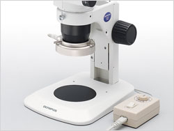
범용 반사/투과광 LED 조명 스탠드는 LED 기술의 모든 장점도 함께 가져다 줍니다. 고르고 밝은 휘도의 LED는 얇은 베이스와 투과 조명의 성공적인 융합을 통해 표본에 대한 접근과 조작이 쉽습니다.
정전기 방전 방지
본체와 주요부속품들은 제전 소재와 코팅의 사용으로 빠르게 정전기를 제거합니다. 이는 관찰 중인 표본을 정전기 손상을 방지합니다.
The STM7 microscopes offer excellent versatility and high performance three axis measurements of parts and electrical components, with sub-micron precision. Whether samples are small or large, simple or complex, or measurements are being taken by a novice or an expert, the Olympus STM7 range features measuring microscopes tailored to fit your needs.
Accurate Measurements through the Integration of an Optical Microscope and Advanced Measurement Capability
The STM7 was Designed with Emphasis on Ease of Use
Accessories that Widen the Range of Observation and Measurement
Automated Focusing System Provides Superior Repeatability
System for Achieving More Advanced Measurement
Accurate Measurements through the Integration of an Optical Microscope and Advanced Measurement Capability
Observation Performance Refined through Years of Microscope Development
The STM7 series uses the same UIS2 infinity-corrected optical system used in state-of-the-art optical microscopes. As a result, observed images have high resolution and high contrast, with aberration thoroughly eliminated to help ensure highly accurate measurement in minute detail.
Measurement Reliability Enhanced with a Stage-Mounting Plate Crafted from Stone
STM7-LF FEM analysis
To provide further assurance of measurement accuracy, the STM7 series uses a highly durable, vibration-resistant frame with a granite surface plate. As a result of this stability, measurements can be taken at sub-micron-levels while ensuring minimal error.
Continuing to Provide User-friendly, High-Precision, 3-axis Measurement as a Pioneer of Height Measurement
Reflective Active, Confocal Autofocus System Optical Path
As modern manufacturing technology becomes increasingly miniaturized and precise, highly accurate measurements are even more essential—not only along the horizontal XY axes, but also along the Z-axis. Olympus has responded to such needs by being the fi rst to realize an autofocus system for measuring microscopes by means of the reflective active, confocal method.
Dependable Quality Based On a Strict Traceability System
Measuring Microscopes Traceability System
The accuracy of Olympus’ measuring microscopes is controlled by a strict traceability system and Olympus even offers traceable calibration at the time of installation.
The STM7 was Designed with Emphasis on Ease of Use
Offering Stages to Fit the Sample Size at Hand
Common Problems
Short measurement stroke precludes the measurement of larger samples.
Sample rotation required to compensate for shorter Y than X-axis coverage during measurement is time inefficient. Until now, large stages have offered a sufficient measurement coverage on the X-axis, but only less coverage on the Y-axis.
Due to the narrow measurement range, it is impossible to line up large numbers of samples on the stage for measurement at once.
STM7 Solutions
Four types of stages are available, each with a unique square measurement stroke (choose from 50 mm x 50 mm, 100 mm x 100 mm, 200 mm x 200 mm, and 300 mm x 300 mm). From small to large size samples, there is a stage that fits the sample being measured.
A clutch system enables rapid switching between coarse and fine movements. Thanks to this switching function, the stage can also be moved rapidly along the X- and Y-axes, and freely across the XY plane.
The 300 mm square length stage enables the same measurement stroke to apply to both the X and Y-axes, which means it can be used to measure large samples, such as 300 mm wafers and printed circuit boards without changing their orientation.
Use the Same Microscope for Both Low- and High-Magnification Observations
Common Problems
Most conventional measuring microscopes only accept a measuring objective or metallurgical objective, and so are unable to meet the requirements for a wide variety of observations.
STM7 Solutions
The STM7 accepts both a metallurgical objective and a measuring objective by exchanging a revolving nosepiece with a measuring objective adapter. This means that the STM7 combines both metallurgical optics and measuring optics in one microscope. In this way, the STM7 series satisfi es a range of needs, no matter whether measuring a wide area or tiny region, measuring the size of differences betwe en levels, or assisting the user in deciding on the best observation method to choose.
Measuring Objectives
Because the measuring objectives have an extremely long working distance, they provide confidence when focusing on samples with large peaks and troughs while reducing worries of the objective coming into contact with the sample. Furthermore, their low-magnifi cation capability enables wide areas to be observed in a single view.
Metallurgical Objectives
Metallurgical objectives enable high-magnifi cation, highresolution observation capability comparable to that of optical microscopes. What’s more, these objectives can be used not only for brightfield, but also for darkfield and DIC observation.
> Click here for details about UIS2 objective lenses
Manual and Motorized Focusing Model Options
The STM7 Line Includes both Manual and Motorized Focus Options
Focus control is available with either manual or motorized operation. Choose the model that addresses your needs in terms of samples and measurement content, regardless of stage size—with all frames incorporating a linear scale for the Z-axis that enables 3-axis measurement.
Manual Z-axis Focus Models
Manual Z-axis focus models offer excellent cost performance—with familiar handle operation for rapid vertical movement that offers convenience for users who needs to measure samples with variety of heights.
Motorized Z-axis Focus Models
Operability is improved and handling fatigue is reduced for focus and height measurements when using the motorized focus unit. The coaxial knobs for coarse and fine movement offer a feeling similar to manual operation, while the models can also be equipped with an autofocus unit.
A Revolutionary Control Unit Refines Measuring Microscope Usability
Common Problems
Additional functions require additional operational units. Operators can’t always locate the corresponding unit quickly, which significantly reduces measurement efficiency.
Numerous operational units and their power supplies around the main unit occupy valuable working space.
STM7 Solutions
Controllers
With the STM7 series, a single controller makes it possible to perform virtually all measuring microscope operations, including use of readout reset, illumination control, focusing, and autofocus. For efficiency and convenience, the unit can be placed wherever you wish and operated easily with one hand.
Control Box
The power supply and transmission for each unit are combined in a single control box. This preserves maximal work space even when a range of optional functions, such as the focus navigator, are added.
Automatic Light Intensity Adjustment Greatly Improves the Efficiency of Observation and Measurement
Common Problems
Analog volume adjustment used by conventional measuring microscopes does not enable the quantitative assessment of light intensity, which can lead to variability in measured values as light intensity changes.
With conventional measuring microscopes, light intensity may need to be adjusted every time the objective is switched—making for an inefficient workflow.
STM7 Solutions
Close Control through a Quantitative Digital Display of Light Intensity Values
The STM7 series provides a quantitative digital display of light intensity—enabling observations to always be made under consistent illumination conditions.
Light Intensity Manager Eliminates the Need for Manual Adjustment
Light intensity manager can be used with the coded revolving nosepiece configuration. The coded revolving nosepiece automatically detects the switching of objectives. This allows the illumination method and light intensity to be registered for each objective, and adjusted automatically during measurements when the objective is switched. Now there is no need to manually adjust light intensity, which used to be required with every switch between magnifications.
A Detachable Digital Read Out for Preferred Location Enables Swift, Convenient Checking of Measurement Results and Equipment Status
Common Problems
The need to check the operation status of equipment, such as illumination, or measured values on individual units makes overall operation cumbersome.
STM7 Solutions
Digital Indicator Enables the Current Operation Status to be Verified Visually
The indicator displays the device status and settings. The minimum X, Y, and Z-axis values can be switched between 0.1 μm and 1 μm, and the display units can be switched between mm, μm, inches and mil.
Detachable Digital Readout Allows for Individual Preference and Placement
Whether attached to the frame or a desk, the placement of the detachable digital readout is up to the individual user. While standing to take measurements, it can be placed on the side of the frame at almost the same height as the site of observation for an exceptional and easy view. When operating from a sitting position, such as observation or measurements on a monitor via a digital camera or when using the motorized Z-axis focusing model, simply place the digital readout and hand controller on the desk.
Automated Focusing System Provides Superior Repeatability
Achieve Faster, Simpler, More Accurate Height Measurement
Common Problems
When doing visual measurement, variations can arise in the height measurements between different operators. Furthermore, this measurement method is time-consuming and inefficient.
STM7 Solutions
Simple and Highly-Precise Focusing System with Superior Repeatability
The Olympus’ focus navigator delivers highly reproducible height measurement by projecting a pattern within the fi eld of view and identifying vertical deviations. Slight errors can occur in height measurements taken with normal visual observation, even when focus appears to be sharp. The focus navigator, however, enables measurements to be made simply by matching up the marks—thereby reducing operator subjectivity in measurement results.
Focus Navigator
Visual Height Measurement
Autofocus Advantage for Fast and Highly Accurate Height Measurement
Common Problems
During visual measurement, the results of height measurement can vary between different operators.
Manual height measurement requires the operator to repeatedly move the stage and adjust the focus with the handle, making measurement time-consuming and inefficient.
Focusing on minute objects, such as bonding wires, is difficult.
STM7 Solutions
Dedicated Autofocus Unit: Outstanding Reproducibility and Focusing Speed
The STM7 dedicated autofocus unit allows highly accurate height measurements to be made with minimal time, regardless of the level of operator experience. Use of the reflective active, confocal method provides a stable focal point independent of surface roughness or a slanting sample surface, while the small laser diameter enables the use of autofocus, even on minute objects, such as bonding wires.
One-shot Mode
Instantaneously takes autofocus from a roughly focused state to sharp focus located at the center of the field of view.
TRACK Mode
The featured TRACK Mode provides autofocus that tracks the peaks and troughs of the sample, even if the stage is moved, keeping the image continually in focus. This advancement greatly improves the efficiency of Z-axis measurements by enabling observations to be made without taking your hands off the X and Y handles.
Accessories that Widen the Range of Observation and Measurement
Coded Revolving Nosepiece
Combining a coded revolving nosepiece with a digital camera lets you display the objective magnification on-screen during observation and allows you to record that magnification. This convenient feature allows information on your sample and the objective’s magnification to be recorded at the same time when recording a sample.
MM6-EMO/ Erect Image Monocular Tube
Monocular tube for erect images. Can be used in combination with MM6-OCC10x (eyepiece with cross hairs).
STM7-FS/ Foot Switch
Enables hands-free transmission of data, allowing operators to complete measurement without taking hands off the X and Y handles.
SZ-LW61/ White LED Illumination Unit
This light-weight, space-saving design model provides a long operating life and low power consumption. The cost-effective LED illumination unit is also free from the flickering and brightness fluctuation.
SZX2-ILR66+SZX-RHS/ LED Ring Illuminator+Manual Control Unit
SZX-RHS manual control unit enables independent illumination of four-segments of the SZX2-ILR66 reflected LED ring illuminator, which provides clear images with high color temperature. The optimal illumination can be selected from 13 patterns.
Rotatable Stage
Enables easy parallel alignment of sample.
System for Achieving More Advanced Measurement
Measurement Support System
The ability to clearly and easily see the output display component of measuring microscopes is essential. That is why the new Olympus measuring software has been created, helping to deliver complex measurements with greater accuracy. The software also enables the use of digital cameras.
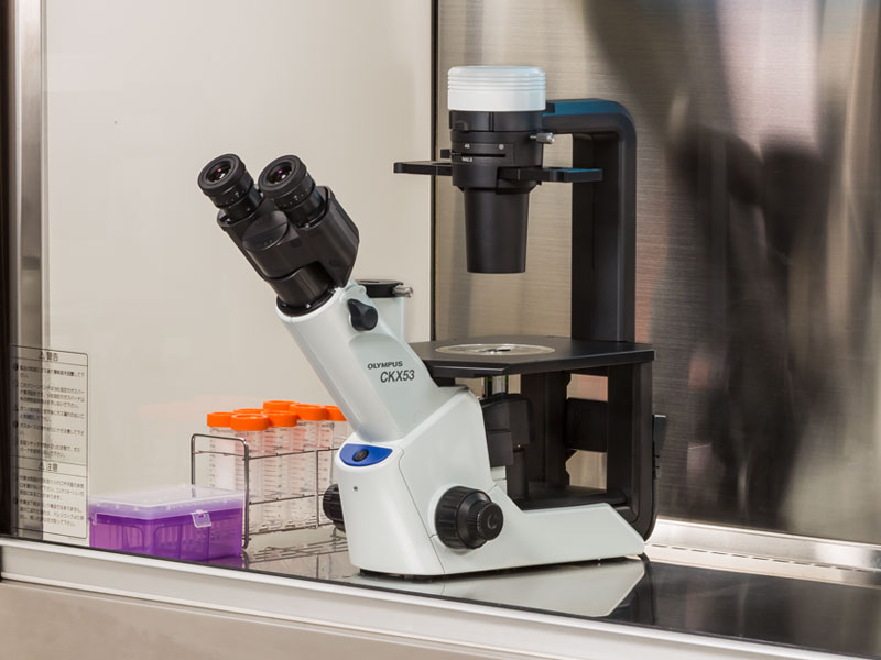
향상된 이미지 품질과 인체공학 설계로, Olympus CKX53은 라이브 셀 관찰, 세포 샘플링 및 처리, 이미지 캡처, 그리고 형광 관찰을 포함한 다양한 세포 배양 샘플에 뛰어난 성능과 효율적인 관찰 흐름을 제공합니다.
라이브 셀 관찰
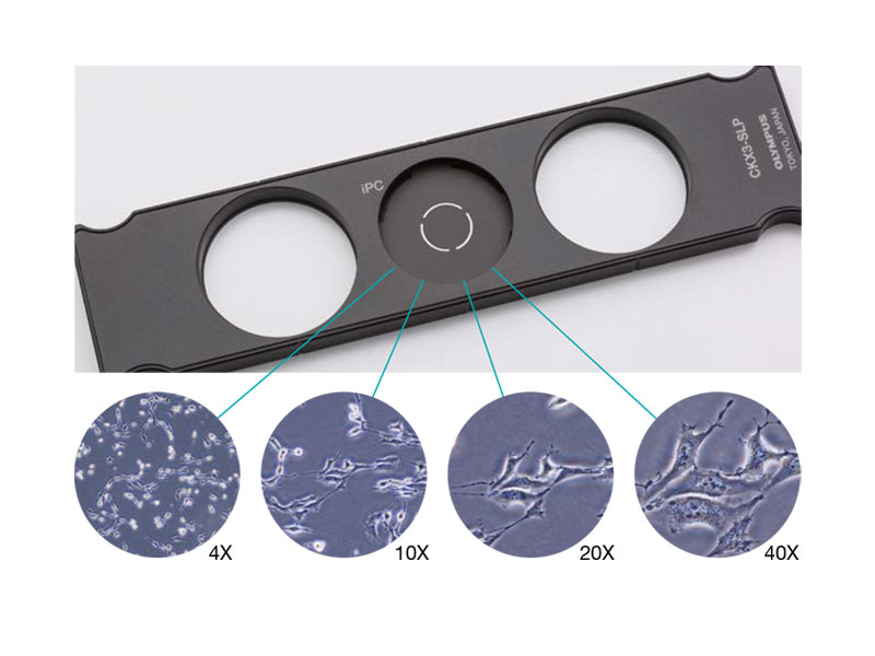
통합 위상차( iPC : Integrated Phase Contrast ) 현미경
CKX53 iPC 시스템을 이용하여 대물렌즈 배율( 4x, 10x, 20x , 40x ) 변경시, 콘덴서 측의 위상차 링슬릿의 연동 변경이 필요 없게 되어 효율적인 관찰 작업이 가능하고, 또한 위상차 설정이 틀어짐에 대한 수시 조정이 필요없어 언제나 선명한 샘플 관찰이 가능합니다.
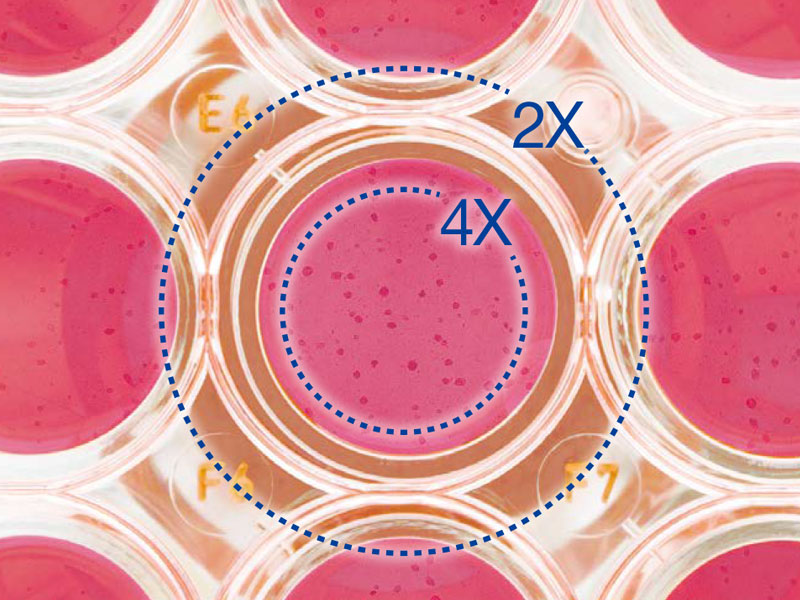
2X 배율, FN 22의 대물렌즈로 선명하고 넓은 시야
PLN2X 대물렌즈를 위한 링 슬릿, CKX3-SLPAS는 직경 11 ㎜ , 시야수 22 ㎜ 를 갖습니다.
2X 대물렌즈는 다른 대물렌즈 보다 확연히 높은 contrast를 제공하여, 투명한 샘플도 명확하게 식별할 수 있습니다. 예를 들어, 96-웰 마이크로 플레이트 관찰시, 넓은 시야로 인하여 스테이지를 움직이지 않고 웰의 모든 세포를 관찰 할 수 있습니다.
IVC (Inversion Contrast) 기술을 사용한 3D 셀 관찰
새로 개발된 IVC 기술로, 위상차보다 시야 심도는 좁아지며, 개체의 모양이나 투명도와 관계없이 삼차원 이미지를 선명하게 합니다. 또한, IVC 관찰은 후광 효과나, 방향성 있는 그림자를 배제하여, 개체의 선명한 관찰을 가능하게 합니다. * 10X 대물렌즈 (PLCN10X, CACHN10XIPC)는 새로운 IVC 관찰에 사용할 수 있습니다.
Glass Heater for microscope
TPi-CKX53X ( Thermo Glass Plate )
Microscope:Olympus CKX53 series
Applicable stage: XY mechanical stage CKX3-MVR
Setting range: ambient ~ 60℃
Plate dimension: W190 x D138㎜
Heating area: W174 × D127㎜
Glass thickness: 0.5 ㎜
형광 관찰 (Fluorescence Microscopy)
다양한 형광 시약과 선명한 시야
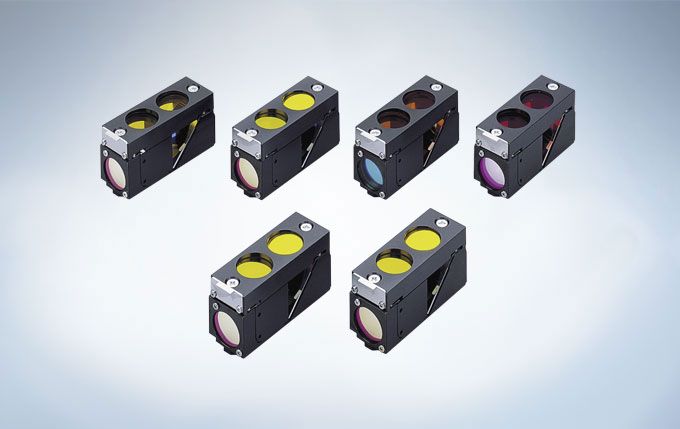
100 W 수은 램프 (U-LH100HG), 130 W 고압 수은 램프 (U-HGLGPS), 그리고 타사(3rd Party) LEDs*와 같은 여러 통합 광원을 이용하여 형광 이미지를 선명하게 관찰할 수 있습니다. 일반 연구용 형광현미경 IX3 및 BX3 에서 사용하는 미러 유닛을 동일하게 사용 할 수 있습니다.
3개의 형광미러 유닛을 장착할 수 있으나, Bright Field 관찰과 위상차 관찰에 영향을 줄 수 있으니 유닛 선택시 고려할 필요가 있습니다.
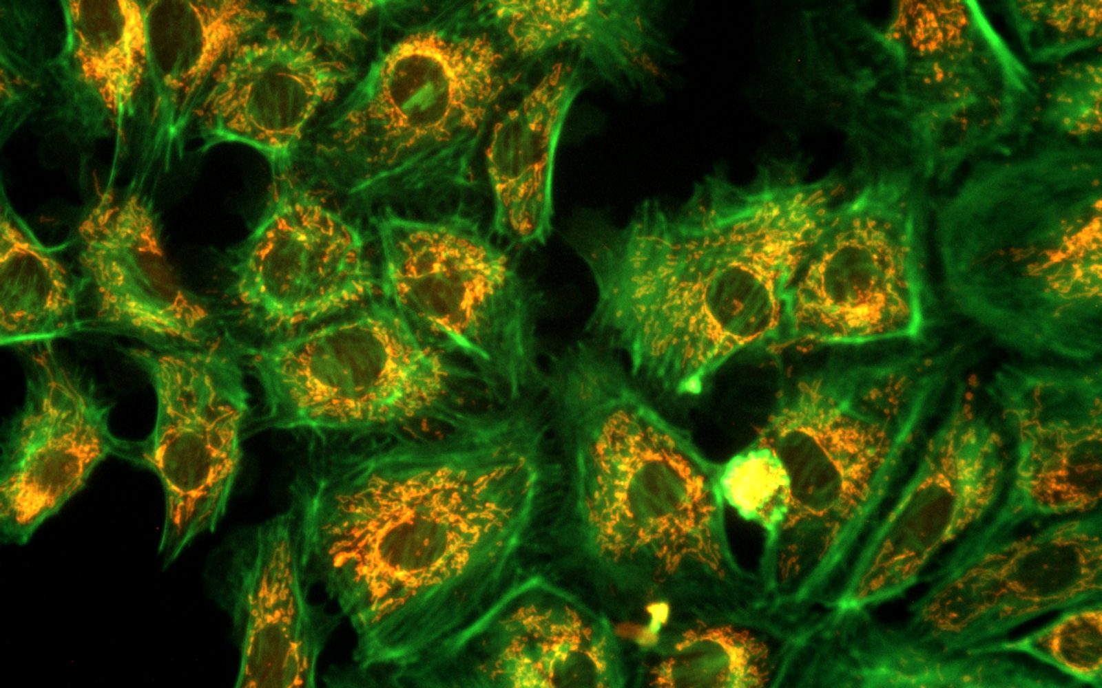
Image taken by AcquCAM 23GR2 with LUCPlanFLN40x Ph2, 1x Adapter, CKX53
밝은 조건에서 높은 Contrast
“Umbra Shield”는 특히 CKX53을 사용한 형광 관찰을 위해 설계되었습니다. 차단막은 실내 광원을 효과적으로 차단하여 형광의 대비를 향상하여 밝은 실험실 조건에서도 선명한 형광 관찰이 가능합니다. 위상차를 사용하는 경우, Umbra 차단막을 들어 올려 표본에 빛을 통과시킬 수 있습니다.
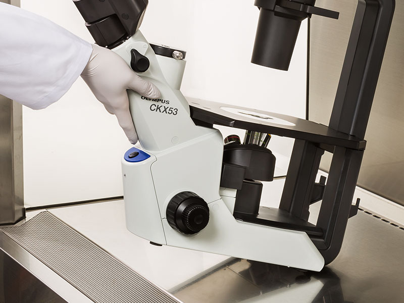
CKX53 현미경은 UV 차단 코팅 덕분에 UV 살균 공정 중에 그대로 둘 수 있습니다. 이 시스템은 약 7kg (15.4lb)으로 이전 모델보다 가볍고 설치 공간이 더 작기 때문에 실험실 공간을 덜 차지합니다. 또한, 한 손으로 현미경을 움직일 수 있으며 관찰 경통의 목 부분을 이용하여 쉽게 운반 할 수 있습니다.
멸균 벤치 환경에서 간편한 세포 샘플링
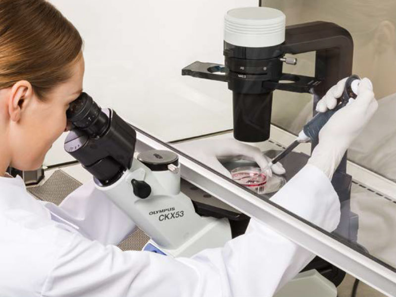
CKX53의 접안렌즈와 광축/포커스 노브 사이의 거리가 짧으므로 작업자의 손의 위치를 자연스럽게 잡을 수 있어서 초점 및 셀 샘플링이 용이합니다.
다양한 세포 배양 용기를 사용할 수 있습니다.
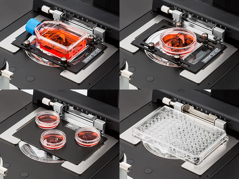
CKX53의 공용 홀더로, 디쉬, 마이크로플레이트, 플라스크를 포함한 다양한 용기에서 배양된 세포를 확인하기 쉽습니다. 옵션 홀더가 부착되면, 최대 세 개의 35㎜ 배양 용기를 스테이지에 장착할 수 있습니다. 또한, 다양한 마이크로플레이트를 별도의 홀더 없이 다룰 수 있습니다.
다층 조직 플라스크(Multi-Layer Tissue Flask)를 위한 종합적인 관찰
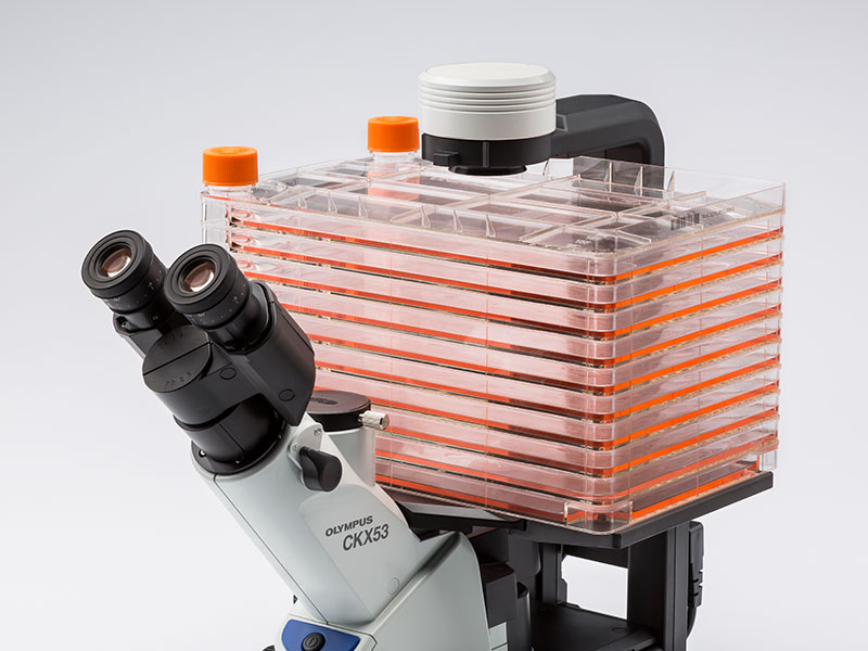
CKX53의 폭과 탈착 가능한 콘덴서로 다층 조직 플라스크와 같은, 최대 190 ㎜ 높이의 배양 용기도 볼 수 있습니다. PLCN4X 대물렌즈의 우수한 초점 심도로 다층 조직 플라스크 내 바닥 두 개의 층의 세포를 빠르고 편하게 관찰할 수 있습니다.
다양한 용기를 사용하여 관찰 유연성 증대
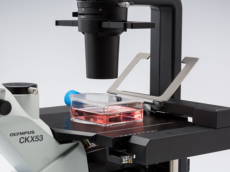
홀더 암을 들어 올려서 수동으로 세포 배양 용기를 배치할 수 있습니다. 또한, 스테이지는 좌우로 최대 70 ㎜ 까지 확장할 수 있습니다.
홀더 암을 들어 올려서 수동으로 세포 배양 용기를 배치할 수 있습니다. 또한, 스테이지는 좌우로 최대 70 ㎜ 까지 확장할 수 있습니다.Product_CKX53_07
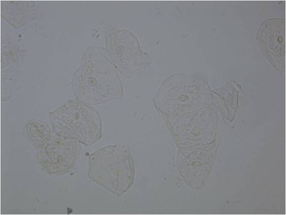
생물현미경에서 사용하는 대부분의 샘플은 무색투명한 특성을 가지고 있기때문에, 배율확대 만을 목적으로 하는 일반 현미경의 관찰법(Bright Field)에서는 투명하고 윤곽이 흐릿하게 보여서 관찰에 어려움이 있습니다.
이 문제를 해결하기 위하여 샘플을 염색하는 방법을 사용하고 있습니다만, 이 방법으로는 염색도주에 살아있는 샘플이 죽어버리기 때문에 살아있는 채로 샘플의 관찰은 할 수 없습니다.
위상차 현미경의 (개요)
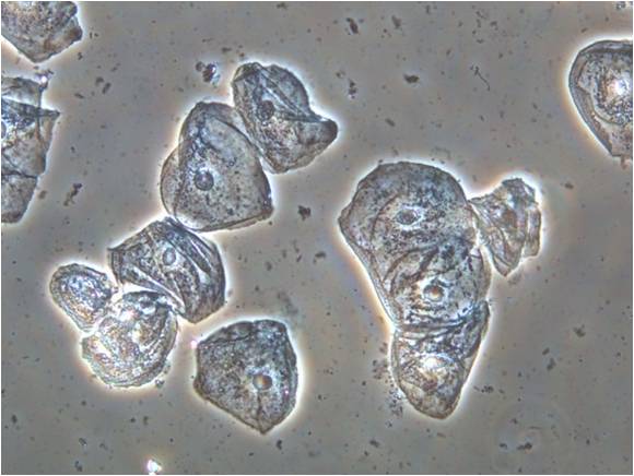
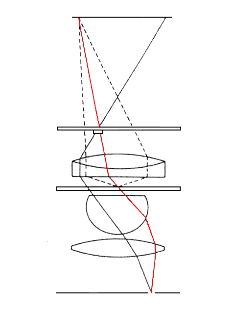
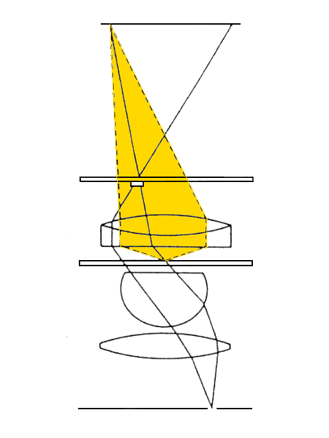
위상차 관찰법(Phase-Contrast)은 샘플을 통과하는 직진광(하단 좌측 이미지)과 이 직진광이 샘플에 통과하면서 발생하는 회절광(하단 우측 이미지) 사이의 위상차 현상을 이용하여 살아있는 세포의 구조와 미생물의 상태변화를 볼게 있게 해주는 관찰법입니다.
위상차 현미경의 원리 설명
샘플의 한 포인트(샘플 평면 중의 한 점)를 통과하는 빛은 두 개의 광학 경로를 가지도록 설계 되어 있으며, 이 두개의 경로를 통과한 빛은 한점에 다시 모여 확대된 상을 만들게 되지만, 다른 경로를 지나왔기 때문에 발생하는 위상차에 의해서 보강 또는 소멸 간섭을 하게 됩니다.
하단의 좌측이미지는 두개의 광학경로 중에 직진광의 경로이며, 하단의 우측이미지는 직진광이 샘플에 닿을때 발생하는 회절광의 경로입니다. 참고로 직진광이 샘플이 없는 포인트를 지나가게 될때는 산란이 생기지않아 회절광은 발생하지 않습니다.
회절광은 직진광의 위상에 비교하여 대략 1/4λ 지연되어 결상하고, 직진광은 위상판에 의하여 1/4λ 또는 3/4λ지연되어 결상하게 됩니다.
직진광이 위상판에 의하여 1/4λ 지연되어 회절광과 동일한 위상을 갖게 되면 직진광과 회절광의 위상이 서로 보강간섭을 하여 진폭이 커지게 되면 배경에서는 직진광의 영향만 받기 때문에 샘플이 배경 보다 밝게 보이게 됩니다. (Negative contrast)
반대로 직진광이 위상판에 의하여 위상이 3/4λ 지연되면 직진광과 회절광은 소멸 간섭을 하게 됩니다만, 다만, 배경(세포가 없는 부분)은 회절광이 발생하지 않기 때문에 직진광에 소멸간섭 현상이 그대로 진행하기 때문에 배경이 더 밝게 보이게 됩니다. (Positive contrast)
별칭 : Normaski Interference Contrast Microscopy ( NIC )
Brightfield Image taken with olympus FV3100DIC Image taken with olympus FV3100Brightfield Image taken with olympus FV3100 (clouseup)DIC Image taken with olympus FV3100 (clouseup) 이미지를 클릭하시면 원본 이미지를 확인하실 수 있습니다.
미분간섭관찰법 발명자
Georges (Jerzy) Nomarski (January 6, 1919 – 1997)
프랑스 귀화 물리학자 (폴란드 태생)
발명 배경
이 관찰법은 샘플이 두꺼운 경우에 후광에 의한 광학 노이즈가 발현되는 위상차 현미경과 다르게 후광 효과가 발현되지 않으며, 샘플에 입체감을 부여하여 투명한 샘플에 대하여 보다 세밀한 관찰을 가능하게 합니다.
이 관찰법은 살아있는 생물학적 시료 및 염색을 하지 않은 조직의 관찰에 폭넓게 사용되고 있습니다.
DIC Microscopy 원리 설명
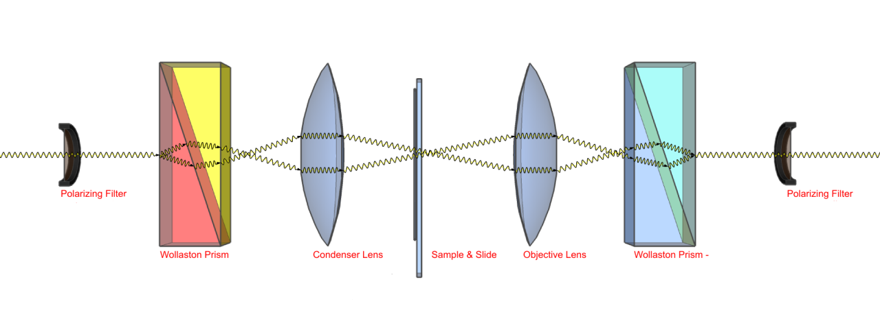
본 관찰법은 편광필터 2개(편광자, 검광자)와 DIC 프리즘 2개의 추가 유닛으로 구성되어 진다.
가장 바깥쪽에 위치하는 편광 필터 2개(편광자,검광자)는 Cross Nichole 상태로 설치 되어 있어야 하며, 이는 두개의 유닛 사이에 광학적 특성의 변화가 있는 빛만을 통과시키겠다는 목적으로 이해하면 된다.
편광자를 통과한 빛이 처음 프리즘에 통과하면 빛은 2개의 경로를 가지게 된다. 이 두 빛은 광학진동 방향이 직교하며, 매우 미소한 거리 차를 가지고 진행한다.
샘플을 통과하고 다음 프리즘을 통과하면서 빛은 다시 동일한 경로를 갖는 빛으로 합성되며, 이때 다른 경로를 통과하였기 때문에 동일한 위상을 갖지 못하는 경우가 발생한다.