J.H.JIN의 모든 글

JNO-DPTS

Multi-Dual Port ( Simultaneously Observation )

Download  Catalog  JNO-DPTS

To observe Fluorescence image  and IR DIC image  at the same time
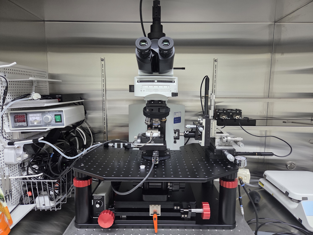
with Multi-Dual Port tube for BX51-WI
(Made by Jae Hwan JIN )

JNO-DPTS JNOPTIC Multi-Dual Port System
  1. Simultaneously Observation
Optical path of JNO-DPTS
Optical path of JNO-DPTS IR-DIC 이미지는 투과 되고, Visivle 이미지 반사되어 각각의 카메라로 전달 된다.

CA3 pyramidal neuron, IR-DIC & Alexa Fluor 488 (50 uM)

1FullF6_2
1FullF6_2
CA3 pyramid neurons taken with JNO-DPTS
CA3 pyramidal neuron Alexa Fluor 488, IR-DIC (Simultaneous observations)
Figure courtesy by Dr. Sooyun Kim, Seoul National University
CA3 pyramid neurons taken with JNO-DPTS
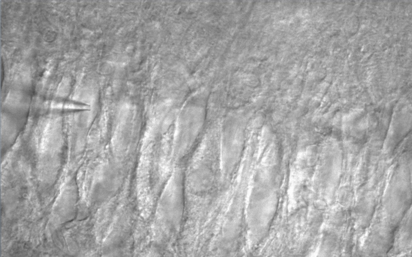
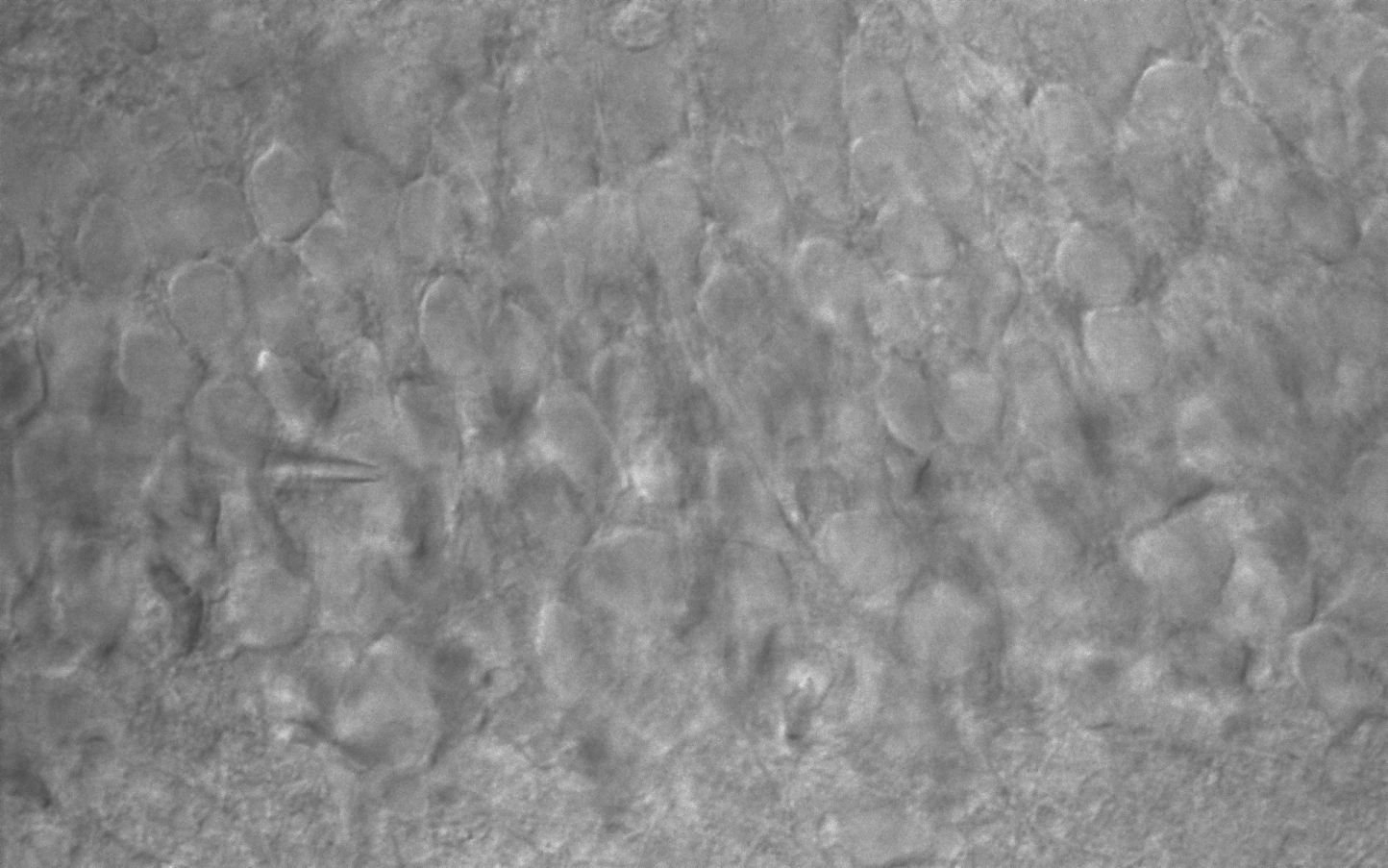
CA3 pyramidal neuron AcquCAM 23S2 & JNO-RNB filter with BX51WI
CA3 pyramid neurons taken with JNO-DPTS
CA3 pyramidal neuron AcquCAM 23S2 & JNO-RNB filter with BX51WI
Frame Rate162 fps, increased through ROI
Sensor1 / 1.2 inch ( 1920×1200, 5.86㎛ x 5.86㎛ )
FILTERJNO-RemN/B (Noisy cut filter)
Manification1x, 1.25x, 1.6x, 2x

JNO-ARM

Advance Realtime Monitor. Easy-to-Use, Image Enhancement, etc

1. Information

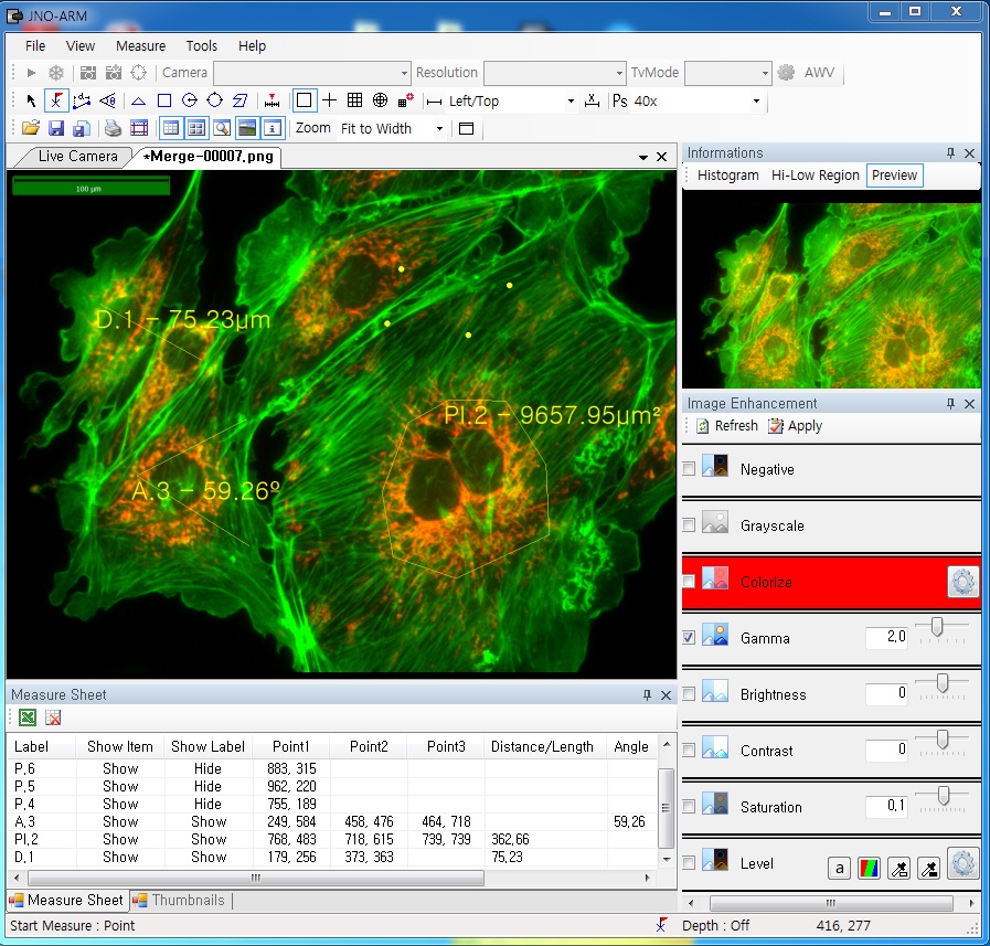
ARM is image analysis software for JNOPTIC AcquCAM cameras. This S/W is interchangeable with all WDM cameras regardless of camera brand and model and even more the most strength point is simple and easy use of length, area, angle, etc.ARM01
2. Simple measurement tools

toolbar

  • Measurement Tools: Count, Distance, Angle, Area, etc.
  • Guide Lines: Cross Line, Rectangular Lattice, Circular Grid
  • Scale Panel on Live/Image Screen
  • Available measurement on LIVE image and saved image mode

3-1. Specialization for observation of fluorescence images

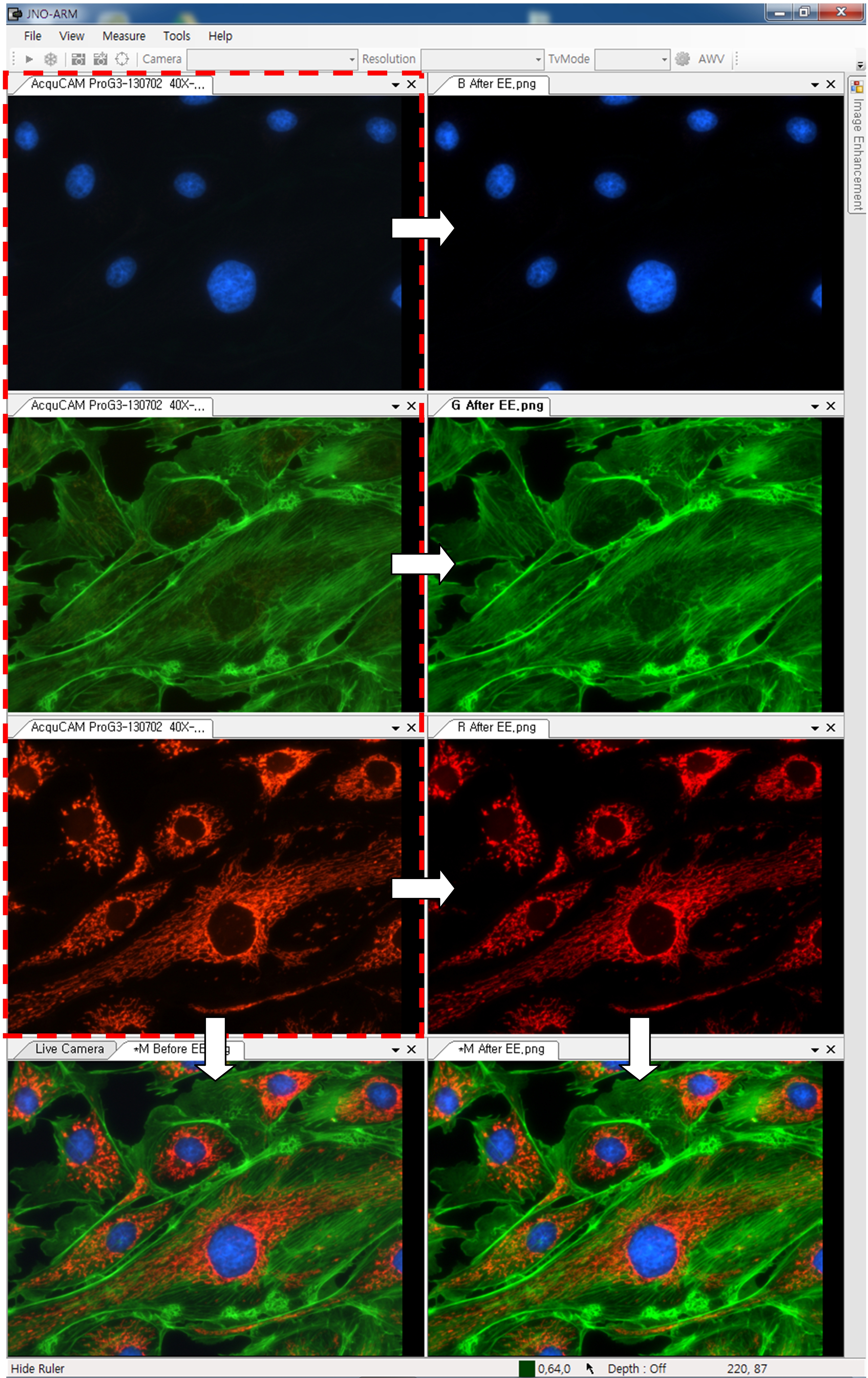
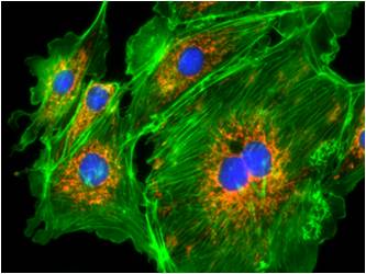
Improvement Function about Fluorescence Microscopy Image. (Used function: Auto Level, Low level, Pseudo Color, Merge Image)
ARM02
 Bottom on the left is merged image without improvement of image. And the bottom on the right is merged image after improvement of image. These images are shot by JNOPTIC AcquCAM Pro/G3 camera

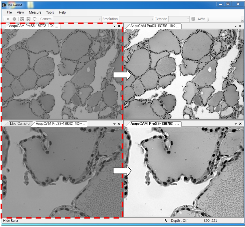
3-2-1 Effect of high-contrast image with simple operation (Monochrome)

Improvement Function about Microscopy Image Monochrome. (Used function: Auto Level)arm04Effective improvement of image with simple operation. This image is shot by JNOPTIC Pro/S3 camera
3-2-2 Effect of high-contrast image with simple operation (Color)

Improvement Function about Microscopy Image. (Used function: Auto Level)arm05Effective improvement of image with simple operation. These images are shot by JNOPTIC AcquCAM Pro/G3 camera
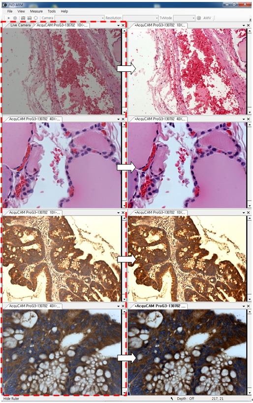
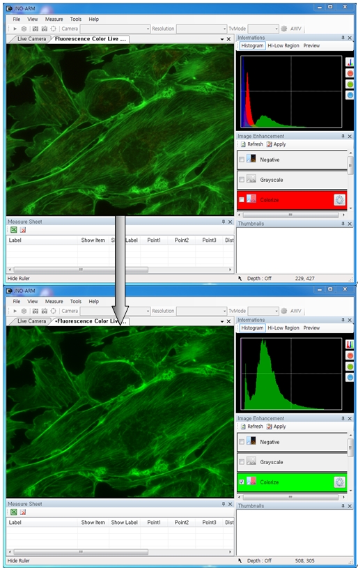
3-3-1 Live Pseudo Color (for color camera)

When you get fluorescence images using color camera, if the unwanted channel was shown because of cross talk, you could remove this channel to take advantage of function of Live Pseudo.
Improvement Function about Fluorescence Microscopy Live Camera Image. (Used function: Live Pseudo Color)
arm06
 The extraction of wanted color information from color image (Remove BR on RGB source) These images are shot by JNOPTIC AcquCAM Pro/G3 camera

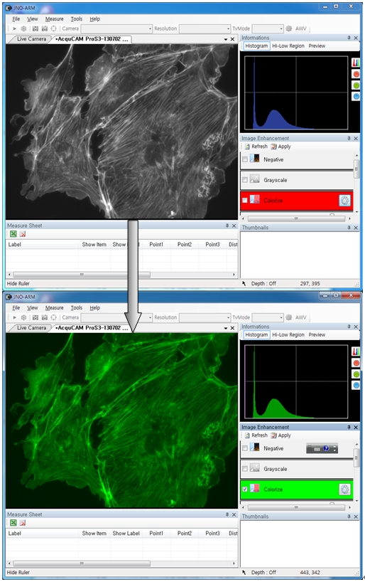
3-3-2 Live Pseudo Color (for Monochrome camera)

When you get fluorescence images using monochrome camera, you can raise efficiency of acquisition for fluorescence images to add similar false color to the color to be shown on the eyepiece in real time.
Improvement Function about Fluorescence Microscopy Live Monochrome Camera Image. (Used function: Live Pseudo Color)
arm08
 Improvement of usage environment for monochrome camera to add false color on the image. These images are shot by JNOPTIC AcquCAM Pro/S3 camera

4. JNO-MHU(Option Unit)

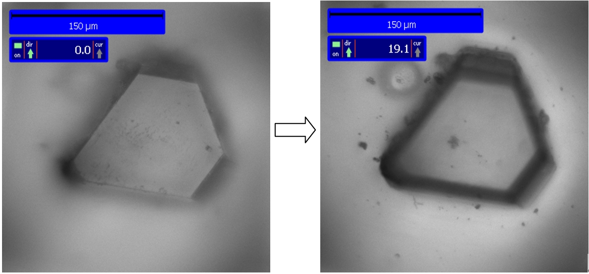
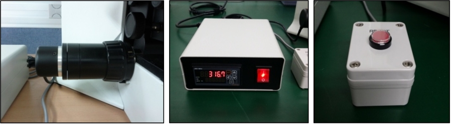
Available measuring height with additional unit, JNO-MHU
※ JNO-MHU is sold separately as optional unit.
jno-mhu1
  jno-mhu2
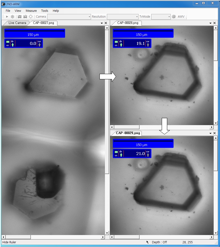
 Left: Focus on top of sample (Z-axis reset), Right: Focus on bottom of sample (Z-axis height measurement)
jno-mhu3
 < Measuring condition : over 20x objective, temp 20°C >
Responding Model Measurement value unit Measurable height Error of measuring 1000㎛ Recommended measuring height
CX 41 0.2 ㎛ -9.9 ~ 29.9㎜ Below ±20㎛ Below ± 2000㎛
CKX 41 0.2 ㎛ -9.9 ~ 29.9㎜ Below ±20㎛ Below ± 2000㎛
BX – FM 0.2 ㎛ -9.9 ~ 29.9㎜ Below ±20㎛ Below ± 2000㎛
BX 51/53 0.1 ㎛ -9.9 ~ 29.9㎜ Below ±10㎛ Below ± 1000㎛
MX 51 0.1 ㎛ -9.9 ~ 29.9㎜ Below ±10㎛ Below ± 1000㎛
MX 61L/61 0.1 ㎛ -9.9 ~ 29.9㎜ Below ±10㎛ Below ± 1000㎛

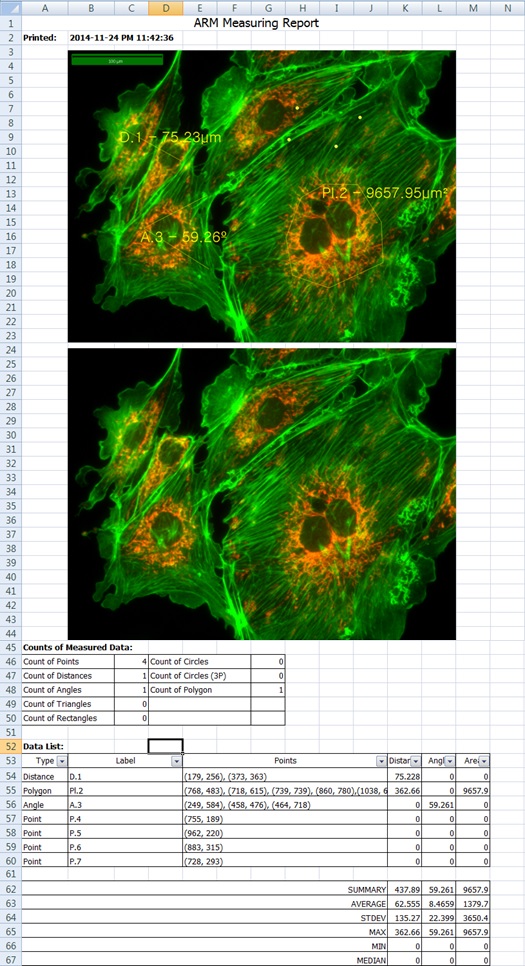
5. Reporting to Excel

excel


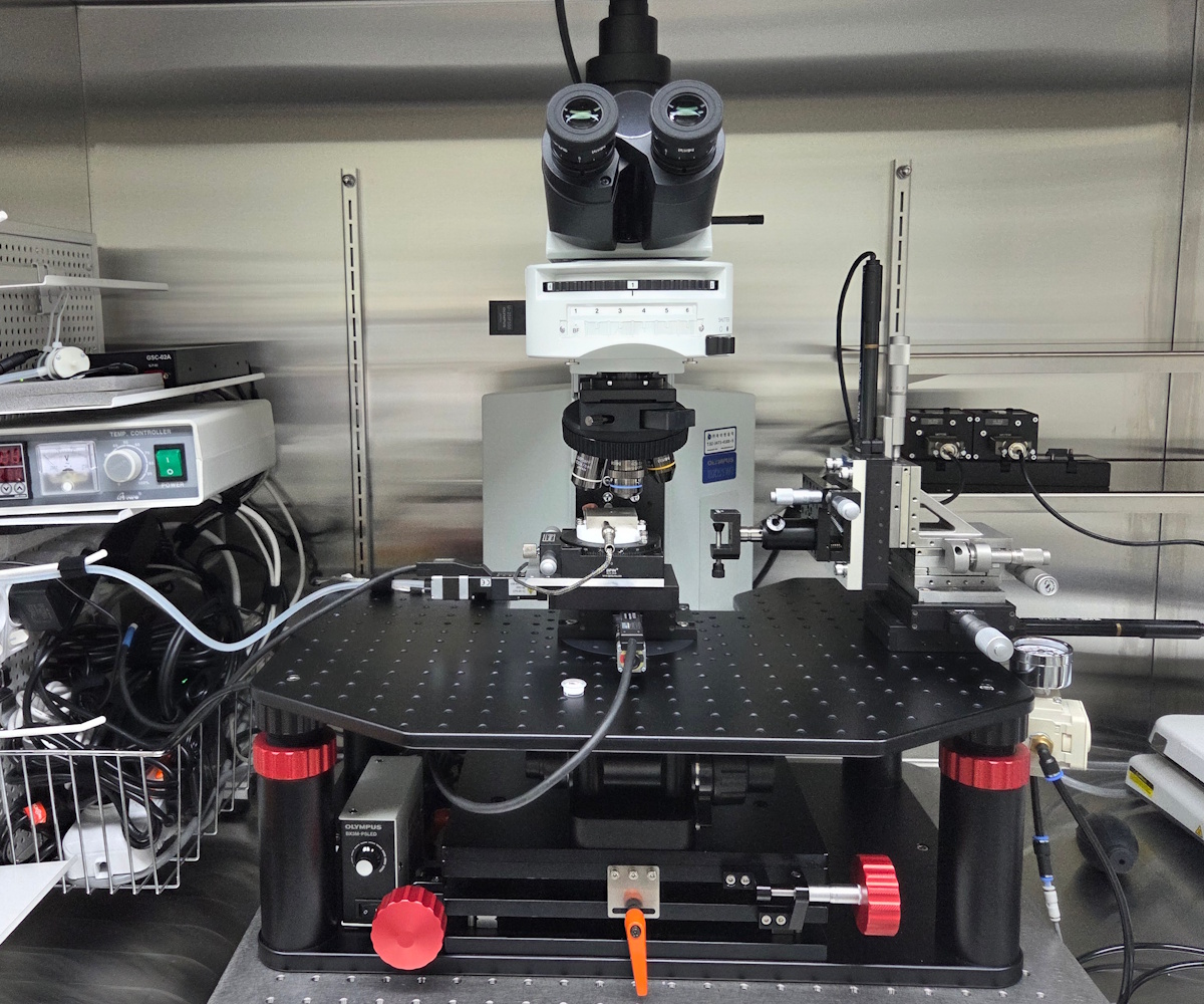
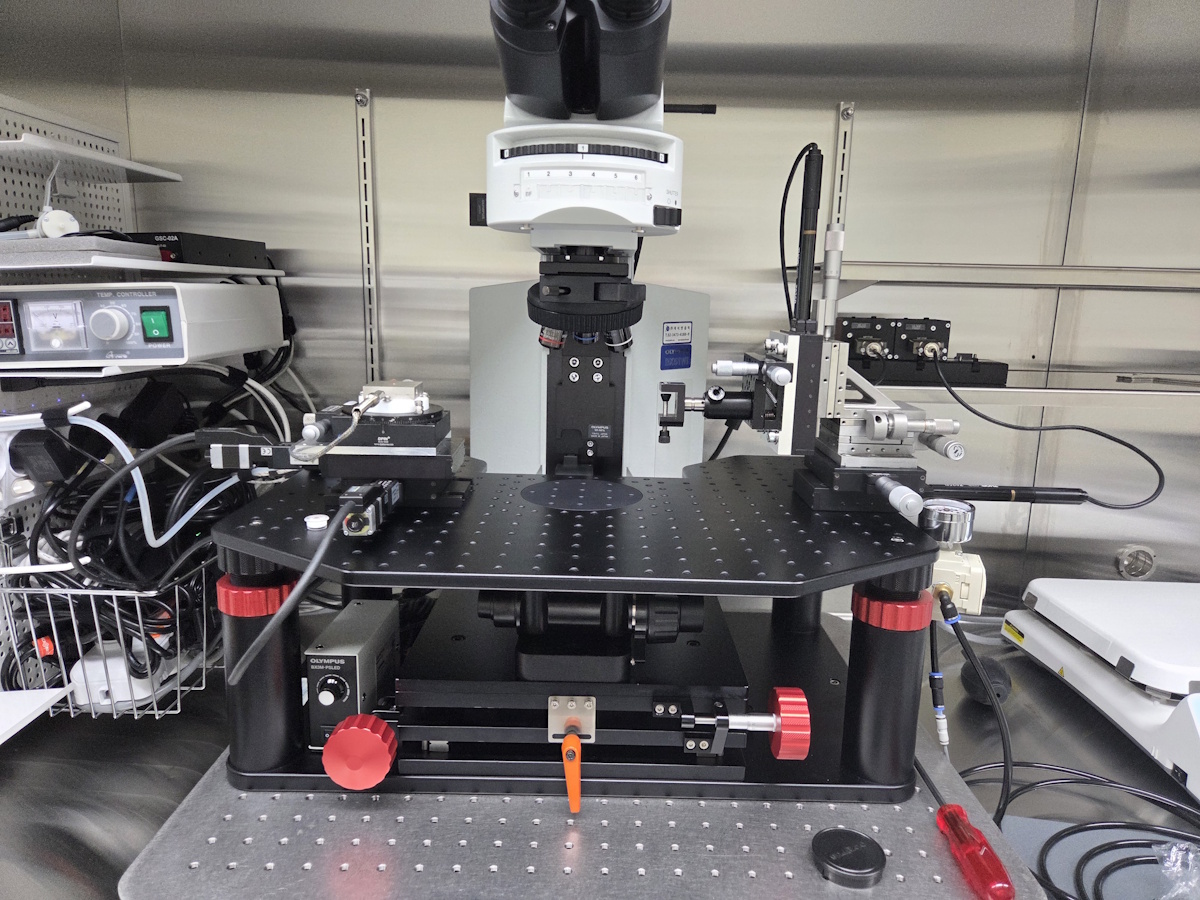
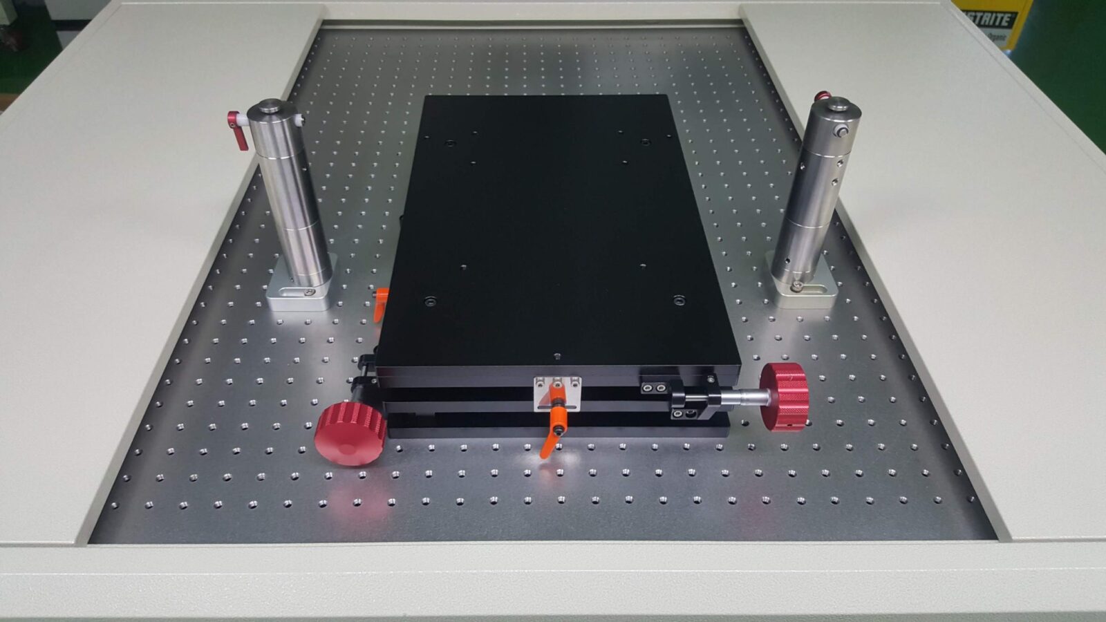
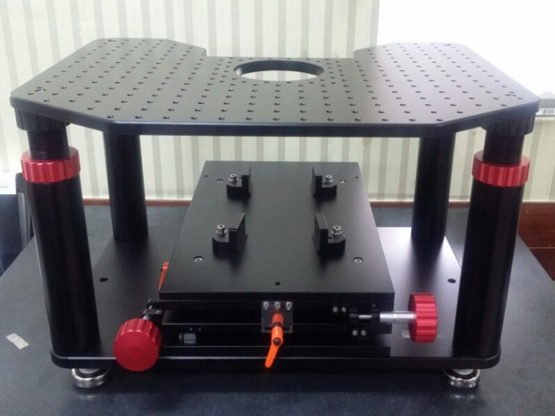
JNO-WI-3

X Y Mover & Bridge Stage

Description
Material  Aluminum
표면 처리  Black –Anodizing
Travel Range (mm)  X    ±12.5     Y    ±12.5
높이 (mm)  170  ~ 220
Tap Hole 규격  M6
Tap Hole 간격 (mm)  X    25    Y    25

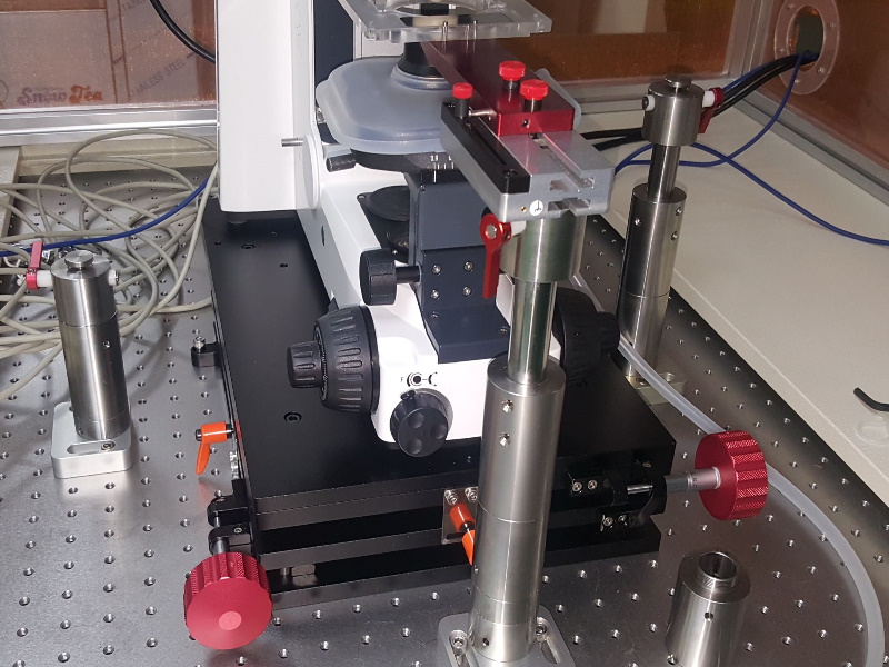
JNO-WI-3 (s)

X Y Mover & Bridge Stage (Short column)

Description
Material  Aluminum
표면 처리  Black –Anodizing
Travel Range (mm)  X    ±12.5     Y    ±12.5
높이 (mm)  100  ~ 150
Tap Hole 규격  M6
Tap Hole 간격 (mm)  X    25    Y    25

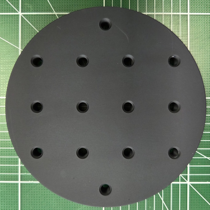
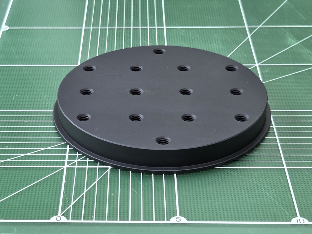
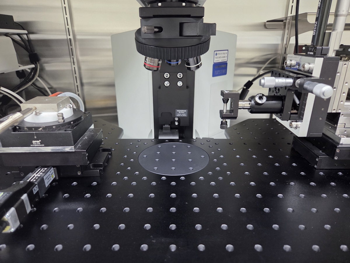
JNO-CP

Centering Plate for JNO-WI-3

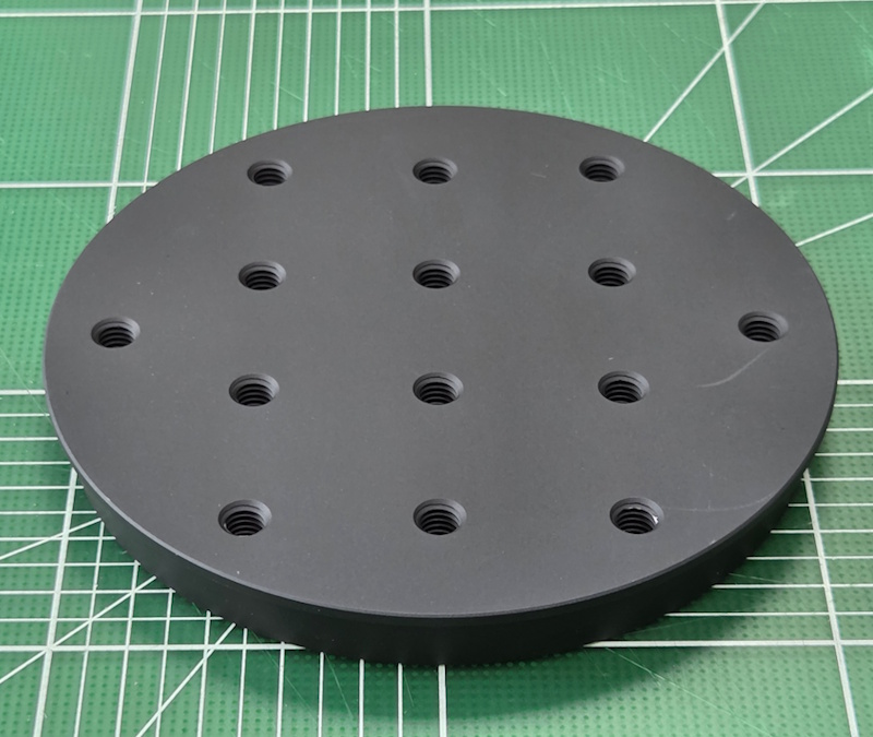
JNO-CP
Centering Plate for JNO-WI-3

XY-MOVER

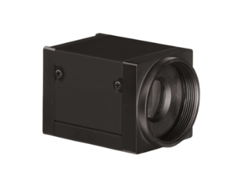
AcquCAM 23S

AcquCAM 23S

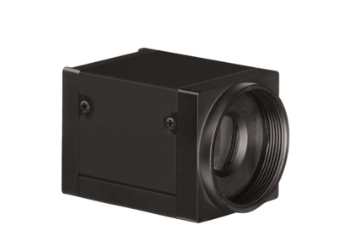
  • USB 3.0 Monochrome Camera
  • 1/1.2 ” Exmor Sensor
  • 1920×1200 pixel
  • Trigger input and I/O

AcquCAM 23S
EXMOR Sensors: Next-generation imaging sensors based on CMOS, increase the strength and reduce noise.


High sensitivity camera available from visible to IR range

20151120_1(CA1 pyramidal neuron)
Apply high sensitivity EXMOR sensor with additional filter block visible range, IR Cut filter image could be observed.


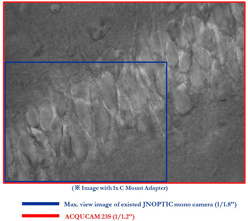
Available wider range of observation

AcquCAM 23S_6
By applying a 1 / 1.2 ” EXMOR sensor, you can observe a wider range of superior sensitivity image.
These images were shot by AcquCAM 23S Package.

High sensitivity and fast frame image

AcquCAM 23S_(CA3 pyramidal neuron)

AcquCAM 23S_(Dentate gyrus Granule cell)
To maximize the range of imaging possible observation with fast frame 54Fps and high sensitivity 0.015 lx .

[Specification]

SpecDescription
Size1/1.2 inch
ResolutionH: 1920, V: 1200
Pixel sizeH: 5.86 µm, V: 5.86 µm
Video formats

& Frame rate

1920×1200 Y16 @ 25 fps

1920×1200 Y800 @ 54 fps

Sensitivity0.015 lx
Exposure time1/100000 to 30 s
Lens mountC/Cs mount
InterfaceUSB 3.0
Power supply4.5 to 5.5 VDC
DimensionsH: 29 mm, W: 29 mm, L: 43 mm
Mass65 g


AcquCAM Pro/S3

AcquCAM Pro/S3

AcquCAM Pro/S3

AcquCAM Pro/S3 USB 3.0 camera is the perfect solution for many bio, industrial, medical applications. Utilizing the very sensitive mono CCD sensors, these cameras feature a specialization of fluorescence observations. With up to 30 fps and a trigger delay of less than 5 micro seconds.

  • 1.2 Mega pixel CCDcamera
  • Monochrome, Progressive scan
  • Preview software provided (JNOPTIC Capture)
  • Software compatible with Windows XP/ Vista/ 7/ 8 operating systems

Apply Pseudo color for live fluorescence

Apply Pseudo color for black live fluorescence

AcquCAM Pro S3_6

Emphasis function for variety images

Getting clear image with general light source

[Specification]

SpecDescription
Size1/3 inch
ResolutionH: 1280, V: 960
Pixel sizeH: 3.75 µm, V: 3.75 µm
Video formats & Frame rate1280×960 Y800 @ 30, 20, 15, 7.5, 3.75 fps 1280×960 Y16 @ 30, 20, 15, 7.5, 3.75 fps
Sensitivity0.015 lx
Exposure time1/10000 to 30 s
Lens mountC/Cs mount
InterfaceUSB 3.0
Power supply4.5 to 5.5 VDC
DimensionsH: 29 mm, W: 29 mm, L: 43 mm
Mass65 g