J.H.JIN의 모든 글

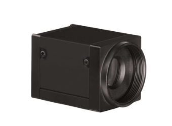
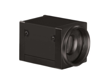
AcquCAM 3

TOMORO AcquCAM 3

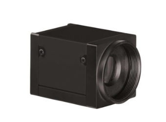
TOMORO ACQUCAM 3는  현미경 영상 획득을 위해 설계되었습니다. 3백만 화소의 이미지 소자는 현미경 영상을 대한 최적화 된 솔루션을 제공합니다. USB2.0 인터페이스를 통하여 하나의 케이블 연결만으로 300만화소의 이미지를 초당 10 프레임으로 관찰 할 수 있으며 최대 35.5 프래임의 고속으로도 현미경 영상을 관찰할 수 있습니다.

Features

  • 3 Mega pixel Micron MT9T031 CMOS Sensor
  • Color, Progressive scan
  • High Speed USB 2.0 Interface (480Mbits/sec)
  • 10  fps at 2560×1536 resolution  /  up to 35.5 fps at 1024×768 resolution
  • Auto White Balance support
  • RGB Bayer video output
  • Hot pluggable
  • All camera parameters adjustable via the computer
  • Preview Software provided (TOMORO Capture)
  • Software compatible with Windows 2K/XP and Vista (32/64bit)  operating systems
  • Fully integrated with OLYMPUS ‘analySIS’ image analysis softare not TWAIN

Specifications

Sensor Micron MT9T031
TypeCMOS Progressive scan
Size1/2 inch
ResolutionH:2560, V:1536
Pixel sizeH:3.2um, V:3.2um
Video formats &(Frame rates)10 Fps at Full resolution
Up to 35.5 Fps at XGA (1024×768) resolution
Sensitivity5 lx at 1/30s, gain 20 dB
Exposure time100us  to 2sec
White balanceAuto / Manual
Lens mountC/Cs mount
InterfaceMini USB 2.0 interface 480Mbit/s
Power supply4.5 to 5.5  VDC via USB 2.0
DimensionsH:50.6mm, W:50.6mm, L:29mm
MassApprox 140g

AcquCAM 카메라 현미경 관찰법 대응표

AcquCAM 카메라 스펙 비교표

Setup Driver & Software Download

AcquCAM Specification Comparison Table

일반 현미경용 카메라

MODEL    No. BKONV
KON3.1
AcquCAM 
3
AcquCAM
5/U3
ConnectUSB 2.0USB 2.0USB 3.0
Sensor CMOS CMOS CMOS
Chip Size 1/2 inch 1/2  inch 1/2.5  inch
Resolution 2048×1536 2048×1536 2592×1944
FPS(Max) 8 10 15
SHUTER rolling rolling
Trigger x x o
Shutter 128㎲ ~ 2s 100㎲ ~ 4s 50㎲ ~ 0.25s
IR Cut 650㎚ ~ 650㎚ ~ 650㎚ ~
Sensitivity 0.05 ㏓
Pixel Size
(µm)
H: 3.2㎛,
V: 3.2㎛
H: 3.2㎛,
V: 3.2㎛
H:2.2㎛,
V:2.2㎛

형광 관찰이 가능한 고감도 카메라

MODEL No. AcquCAM
Pro/G3
AcquCAM
3G
AcquCAM 5GAcquCAM
23GR2
ConnectUSB 3.0USB 3.0USB 3.0USB 3.0
Sensor CCDPregiusPregius Pregius
Chip Size 1/3 inch1/1.8 inch2/3 inch1/1.2 inch
Resolution 1280×9602048×15362448×20481920×1200
FPS(Max) 306035162
SHUTER global globalglobalglobal
Trigger oooo
Shutter 100㎲~ 30s 20 ㎲ ~ 30s 25 ㎲ ~ 4s 20㎲ ~ 30s
IR Cut 650㎚ ~ 650㎚ ~ 650㎚ ~ Option:
700㎚~
Sensitivity 0.05 ㏓0.05 ㏓0.05 ㏓0.05 ㏓
Pixel Size
(µm)
H:3.75㎛, 
V:3.75㎛
H:3.45㎛,
V:3.45㎛
H:3.45㎛,
V:3.45㎛
H:5.86㎛,
V:5.86㎛

전기 생리학 실험 전용 카메라

MODEL    No. AcquCAM 
Silver
AcquCAM 
Pro/S3
AcquCAM 
23S2
ConnectUSB 2.0USB 3.0USB 3.0
TYPEMonoMonoMono
SensorCCDCCDPregius
Chip Size1/1.8 inch1/3 inch1/1.2 inch
Resolution1600×12001280×9601920×1200
FPS(Max)1230162
SHUTERglobalglobalglobal
Triggerxoo
Shutter100㎲ ~ 30s100㎲ ~ 30s20㎲ to 30s
IR CutOption: 700㎚ ~Option: 700㎚ ~Option: 700㎚ ~
Sensitivity 0.05 ㏓ 0.015 ㏓ 0.015 ㏓
Pixel Size
(µm)
H:4.4㎛,
V:4.4㎛
H:3.75㎛,
V:3.75㎛
H:5.86㎛,
V:5.86㎛

AcquCAM 카메라 현미경 관찰법 대응표

Setup Driver & Software Download

Image-Pro v11

Image-Pro logo

ImagePro11 Feature Introduction

File Management, 2D Viewer, Process/Adjust, Usability, Data Management, Reports, Auditing, Scripting

Direct & Assisted Measurements, Auto Shape & Edge Detection, Classification, & Color Measurement.

Smart Segmentation, Classification, 2D Tracking, & the Essentials Protocol Collection with 3 Protocols.

Object CountPercent Area
Objects within Objects
Includes 3 Protocols
상기 테이블의 기능은 하기의 동영상에서 확인하실 수 있습니다.
ApoptosisAutophagy
Cell CountCell Morphology
Cell ProliferationConfluence
Lipid DropletLive/Dead
RingTransfection 1 Label
Transfection 2 Label
Includes 10 Protocols
상기 테이블의 기능은 하기의 동영상에서 확인하실 수 있습니다.

ApoptosisAutophagy

Cell CountCell Morphology

Cell ProliferationConfluence

Lipid DropletLive/Dead
RingTransfection 1 Label
Transfection 2 Label
AngiogenesisColocalization
Neurite OutgrowthTranslocation Total Cellular Area
Wound HealingTranslocation Area Above Threshold
Includes 5 Protocols
상기 테이블의 기능은 하기의 동영상에서 확인하실 수 있습니다.

AngiogenesisColocalization

Neurite OutgrowthTranslocation Total Cellular Area

Wound HealingTranslocation Area Above Threshold
Composite MaterialsLayers Concentric
Layers CurvedLayers Parallel
Fiber SeparationFiber Thickness
Particles SizeParticles Phase
Phase Pores
Includes 10 Protocols
상기 테이블의 기능은 하기의 동영상에서 확인하실 수 있습니다.

Composite MaterialsLayers Concentric

Layers CurvedLayers Parallel

Fiber SeparationFiber Thickness

Particles SizeParticles Phase

PhasePores

ImagePro11 Compared to ImagePro10

Image-Pro logo

Image & MetaData Management

  • Channel Management – Save unique characteristics per channel

Views & Overlays

  • Well Plate Viewer
  • Color-Coded Well Plate Heat Maps

Search & Help

  • Universal Search across Recently Used items & Help topics
  • Dockable & Dynamic Help panel for easy access to Documentation

Licensing

  • Flexible License Linking/Unlinking to Computer or License Server in addition to USB dongles
  • Online License “Bundle” management – Control module configuration per base product

Data Collection & Display

  • Display Data using Heat Maps
  • Display annotated images in Data Table along with statistical data

Classification

  • Adjust measurement overlay appearances by class

Protocol-based Segmentation

  • Run on single file multi-positional (including multi-well) datasets or collections of individual images
  • Bi-direction feedback between data tables & annotated images & charts
  • Library of Protocol reports for both large datasets & single images
  • Analysis Protocols – Essentials Collection (Objects within Objects, Object Count, % Area)
  • Analysis Protocols – Cell Biology Collection (Apoptosis, Autophagy, Cell Count, Cell Morphology & more)
  • Analysis Protocols – Cell Biology Advanced Collection (Angiogenesis, Colocalization, Neurite Outgrowth, & more)
  • Analysis Protocols – Materials Collection (Particles, Pores, Particle Phase, Fiber Thickness, & more)

Multi-Channel Capture

  • LightSource Device Control
  • Acquisition of mixed multi-channel & transmitted light datasets
  • Automated capture of multichannel image-set (w/ a supported multi-channel light source)
  • Guided acquisition of multi-channel image-sets from a manual microscope

Real-Time Deconvolution

  • 2D Real-Time Deconvolution integrated with Capture
  • Wiener Filter-based algorithm
  • 2D Iterative-based algorithm

3D Visualization

  • 3D viewer with advanced control & volume rendering (Min & Max Projections)
  • Clipping plane viewing
  • Slicer viewing

3D Visualization of Big Data Sets

  • Convert Large Image Sets Into Pyramidal Tiff for Big Data Visualization
  • 2D Fixed & Adaptive Deconvolution Algorithms
  • Support of both Theoretical & Measured PSFs
  • GPU Acceleration
  • PSF generation for WF, CF, 2 Photon, Spinning Disk, & STED modalities
  • Spherical Abberation Correction
  • Batch Deconvolution
  • Bundles can be left as floating licenses or can be fixed to a single workstation
  • Bundle assignment to License Server can be adjusted anytime as needed
  • User time out returns license back to License Server

JNO-ICG

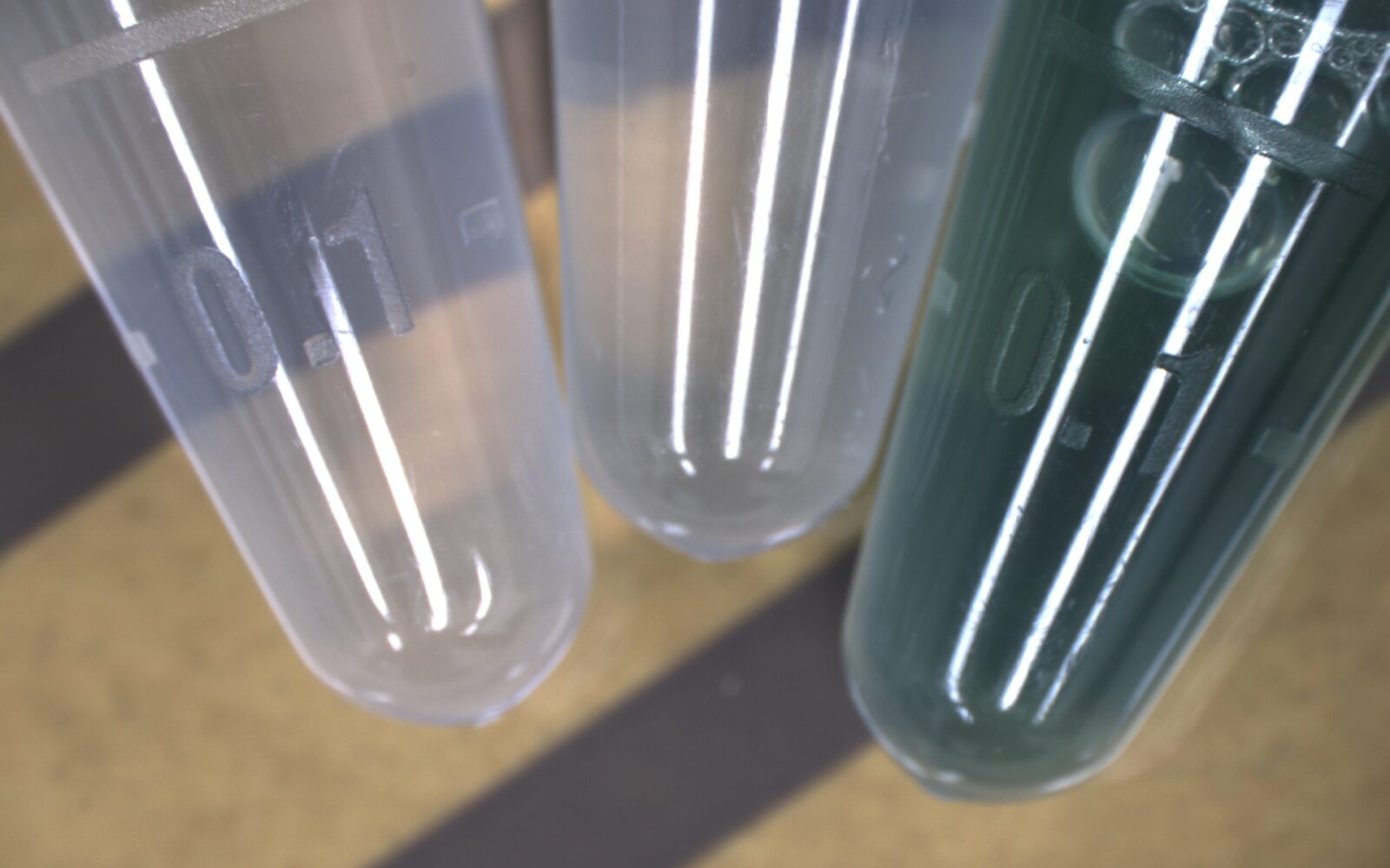
Indocyanine Green (ICG) 관찰을 위한 실체 현미경

Stereo-microscope for Indocyanine Green

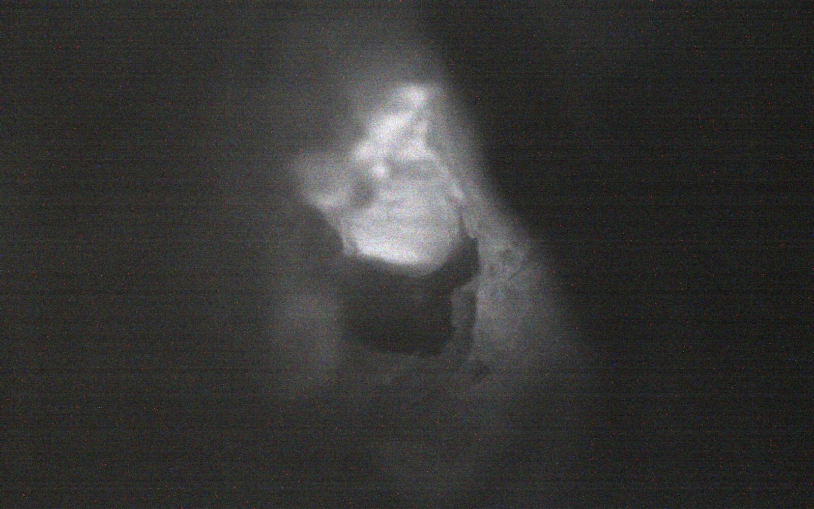
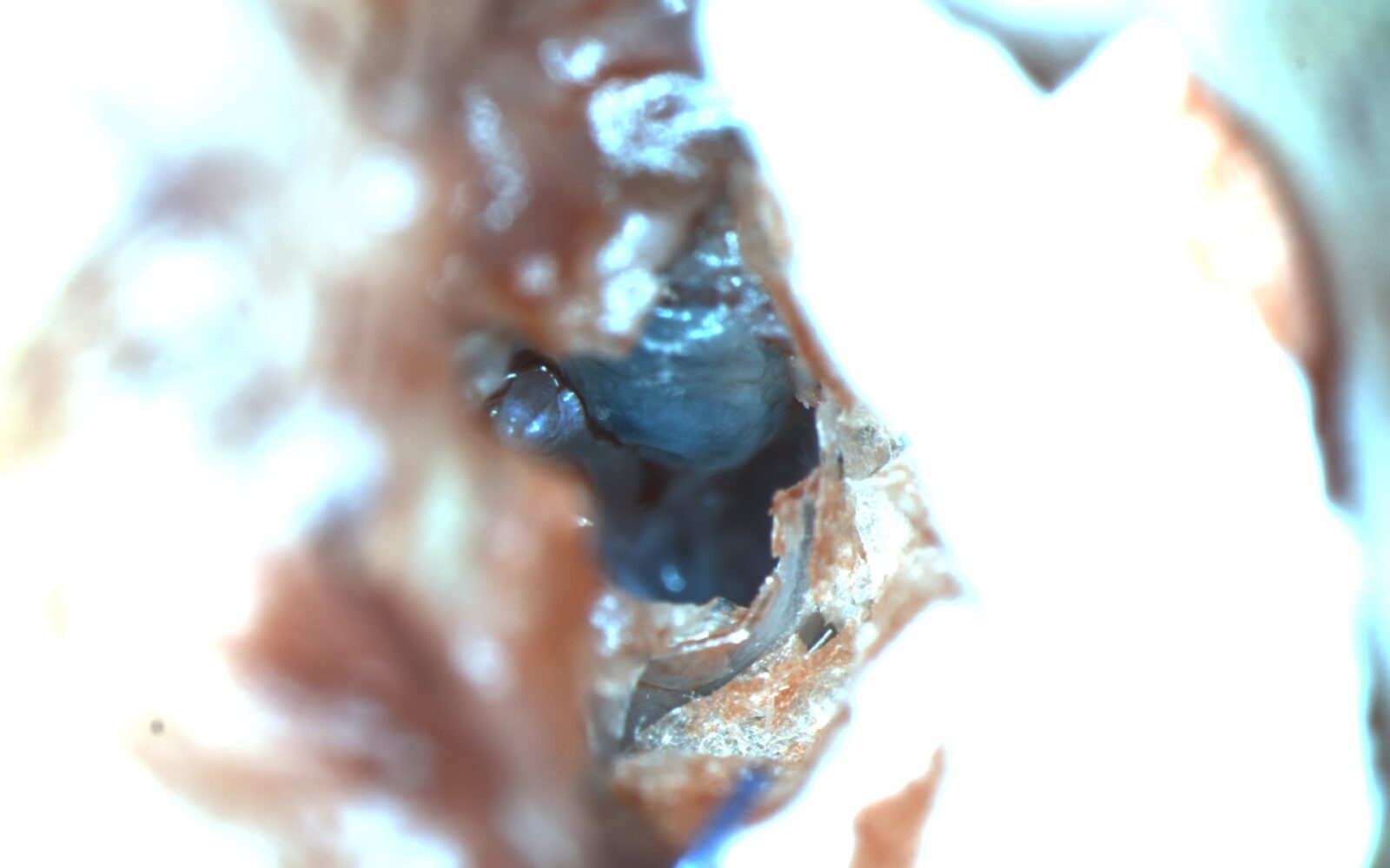
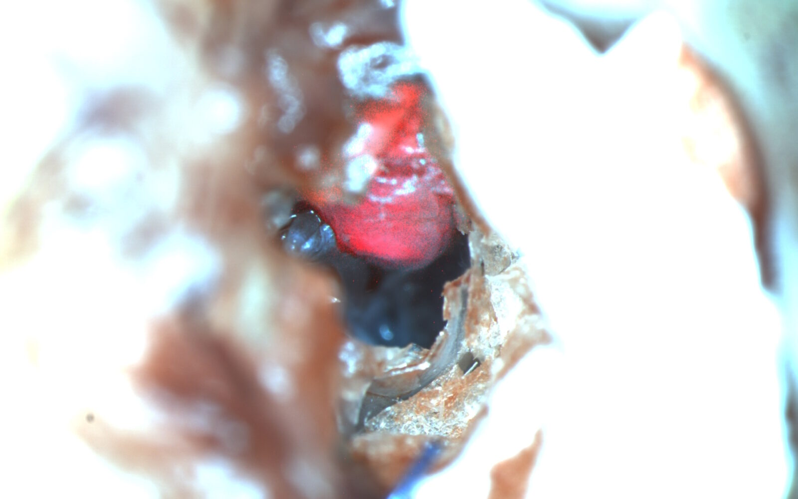
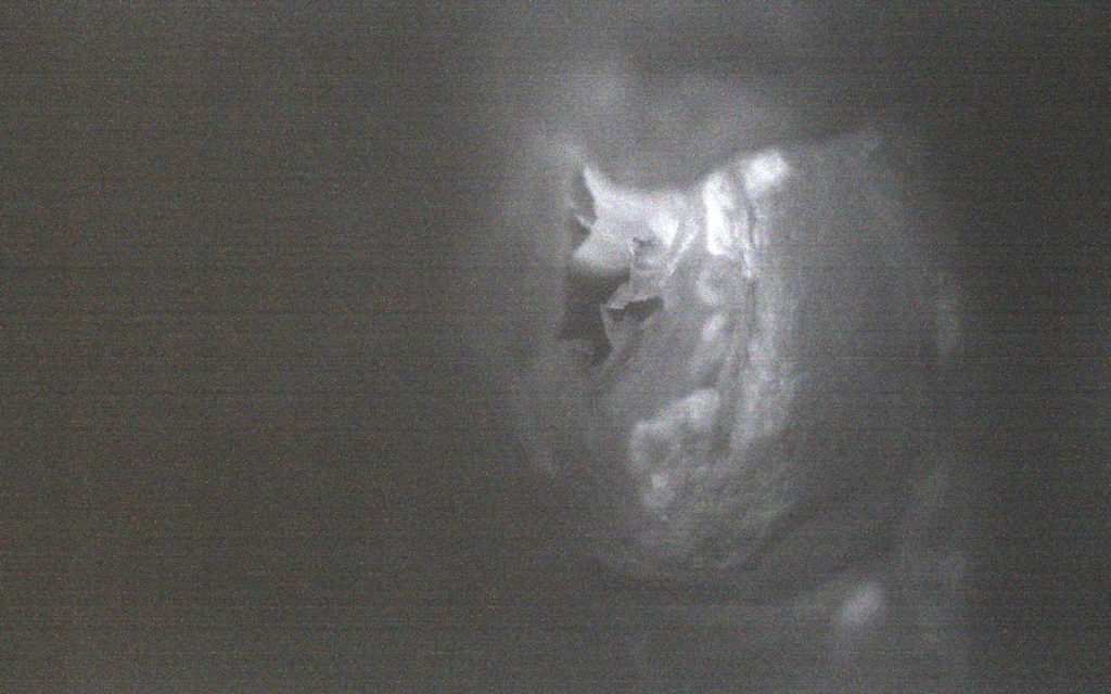
Images captured with JNO-ICG

Indocyanine Green (ICG) _ Bright Field ImageIndocyanine Green (ICG) _ Fluorescent & Bright Field Image
Indocyanine Green (ICG)
Fluorescent & Bright Field Image
Indocyanine Green (ICG) _ Bright Field ImageIndocyanine Green (ICG) _ Fluorescent & Bright Field Image
Indocyanine Green (ICG)
Fluorescent & Bright Field Image
Indocyanine Green (ICG) _ Fluorescent ImageIndocyanine Green (ICG) _ Bright Field Image
Indocyanine Green (ICG)
Fluorescent & Bright Field Image
  • JNO-ICG에 관련한 문의는 하기의 담당자에게 연락바랍니다.
E-mail : jhjin@jnoptic.com 
Mobile : + 82-2-3473-4188~9

Fluorochrome – Cy7™

Cy7™

Spectrum Chart

Cy7 Spectrum
Excitation (㎚)Emission (㎚)
743767

Recommended Filter set

  • 일반 형광 Lighting source
Model No.ExcitationDichroic mirrorEmissionRemarks
JNO-49007ET710/75xT760lpxrET810/90m
JNO-49037ET740/40xT770lpxrET780lp

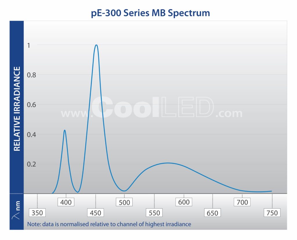
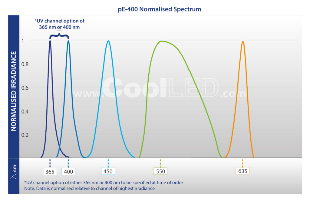
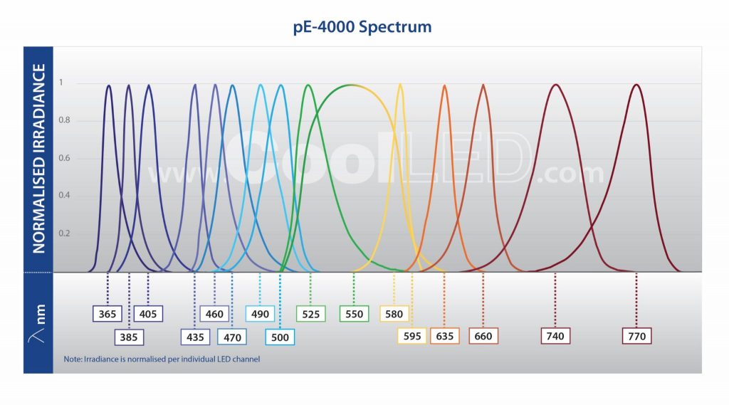
Cy7 & JNO-49007 Specrtum Chart for CoollED pE4000
Cy7 & JNO-49037 Specrtum Chart for CoollED pE4000
  • Cy7을 관찰하기 위한 형광 광원 장치
    • T. 02-3473-4188로 연락 주시기 바랍니다.


Information

시아닌은 여전히 업계에서, 그리고 최근에는 생명 공학 (라벨링, 분석)에서 사용되고 있습니다. 시아닌은 형광 염료로서, 특히 생체 의학 이미징에서 많은 용도로 사용되며 구조에 따라 IR에서 UV까지 스펙트럼을 커버합니다.

Cyanine은 polymethine 그룹에 속하는 합성 염료 계열의 비 계통적 이름입니다. cyanin이라는 단어는 일반적으로 청록색 ( “아쿠아”에 가까운)의 그늘을 의미하는 영어 단어 “cyan”에서 유래되었으며 다소 다른 색상 “진한 파란색 “을 의미하는 그리스어 κυάνεος / κυανοῦς kyaneos / kyanous에서 파생되었습니다.

Cy7은 육안으로는 보이지 않는 근적외선 형광체이며, Cy7.5 염료와 함께 생체 내 이미징 어플리케이션에 사용됩니다.

Cyanines (Cy)는 Fluorescein (FITC) 및 rhodamines (TRITC, RRX)와 같은 기존의 염료를 대체하여 보다 밝고 안정한 형광을 생성합니다.

Cyanines were and are still used in industry, and more recently in biotechnology (labeling, analysis). Cyanines have many uses as fluorescent dyes, particularly in biomedical imaging. Depending on the structure, they cover the spectrum from IR to UV.

Cyanine is the non-systematic name of a synthetic dye family belonging to polymethine group. The word cyanin is from the English word “cyan”, which conventionally means a shade of blue-green (close to “aqua”) and is derived from the Greek κυάνεος/κυανοῦς kyaneos/kyanous which means a somewhat different color: “dark blue”.

Cy7 is a near-IR fluor that is invisible to the naked eye (Excitation/emission maximum 750/776 ㎚). It is used in in vivo imaging applications, as well as the Cy7.5 dye.

Cyanines(Cy) replace advantageously conventional dyes such as Fluorescein(FITC) and rhodamines (TRITC, RRX), yielding brighter and more stable fluorescence.

LIGHT SOURCE FOR FLUORESCENCE MICROSCOPE

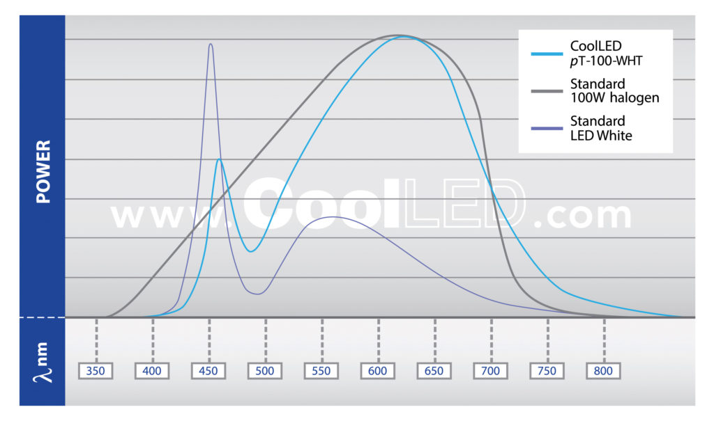
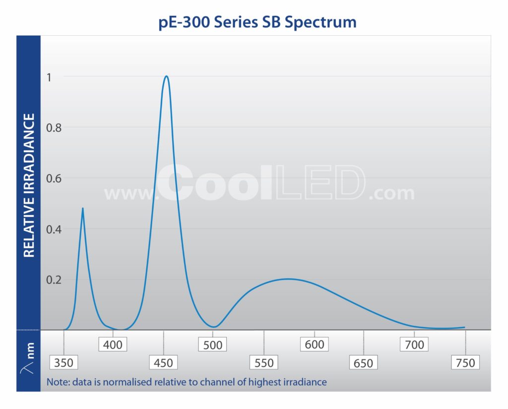
CoolLED pE-300lite

  • 수명 :  25,000시간
  • UV (DAPI) ~ Red (Cy5 EX) 의 스펙트럼
  • 무수은 대체 장치
  • 1 % 단위의 정밀한 밝기 제어 (0-100 %)

CoolLED pE-300whtie

  • 수명 :  25,000시간
  • UV (DAPI) ~ Red (Cy5 EX) 의 스펙트럼
  • 무수은 대체 장치
  • 1 % 단위의 정밀한 밝기 제어 (0-100 %)
  • RGB 3채널 밝기 개별 제어
  • TTL & USB Control

CoolLED pE-300ultra

  • 수명 :  25,000시간
  • 파장 강도 및 셔터를 정밀하게 제어 가능
  • 다중 TTL 입력
  • 필터큐브 변경 없음
  • Sequence runner

CoolLED pE-340fura

  • 수명 :  25,000시간
  • Mercury-free
  • Energy Efficient: 80% less power
  • 빠른 리모트 컨트롤
  • Sequence runner

CoolLED pE-4000

  • 수명 :  25,000시간
  • 365~770nm 파장대
  • Instant on/off – no shutters
  • TTL & USB interfaces
  • Mercury-free & Laser-free
  • 수은 램프 교체 주기 :  평균 2,000시간
  • Powerful 130 W burner with long life time
  • Stable and uniform light output

U-LH100HG

  • 수은 램프 교체 주기 :   대략 200시간
  • 100 W Mercury Light Source
  • Entry-level fluorescence excitation light source
  • Applicable for all common fluorophores

U-LH75XEAPO

  • 제논 램프 교체 주기 :   대략 200시간
  • 75 W xenon burner
  • Ideally suited for far-red and IR excitation
  • Highly corrected for optical

U-LH50HG

  • 수은 램프 교체 주기 :   대략 200시간
  • 50 W Mercury Light Source
  • Entry-level fluorescence excitation light source
  • Applicable for all common

USED-MICROSCOPE

중고 및 데모 현미경 판매의 건

중고 장비 필요하신 분은 하단의 연락처로 연락 바랍니다.

주소: 서울시 서초구 남부순환로 335길 35 일광빌딩 201호
T. 02-3471-4188~9, E-mail : jhjin@jnoptic.com
담당자 : (주) 제이엔옵틱 진재환 올림


생물 현미경
( Bio-Microscope )

투과 현미경 ( 옵션 : 반사 조명 관찰 추가 )
대물렌즈 : 4x 10x 40x 100x PlanC
샘플 높이 측정 기능 ( 최소단위 : 0.2 ㎛ )
카메라 부착 가능

구형 생물 현미경
( Oly type Bio-Microscope )

CHB_OLYMPUS_Used-Microsocpe
생물 현미경 (Old Type Bio-Microscope)
대물렌즈 : 10x 40x 100x
(110V 전용)