J.H.JIN의 모든 글

PE-4000


CoolLED pE-4000


Overview

NEW GENERATION LEDs The pE-4000 sets the standard as the universal illumination system for fluorescence microscopy.  The system has 16 selectable LED wavelengths across 4 channels that can be matched to the filters and fluorophores of almost any microscope, making it the broadest spectrum of illumination available.

The CoolLED pE-4000 now benefits from our award winning sustainable Greentechnology and delivers enhanced intensity at your microscope sample plane with a significant reduction in energy consumption, and is supplied with a 3 year warranty after registration.
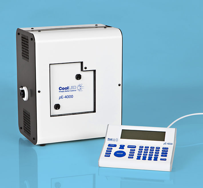
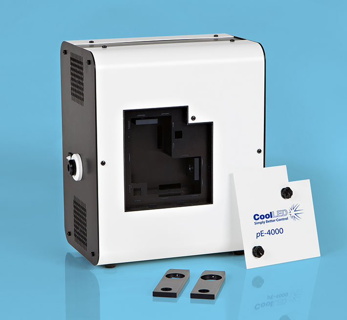
 

The NEW Enhanced pE-4000 is a powerful 4-channel high-specification LED illumination system. It is flexible, controllable and environmentally friendly.  Benefits include:

SELECTABILITY

  • Choice of 256 wavelength combinations from 16 installed LEDs (365nm -770nm)
  • Choose 1 of 4 wavelengths for each 4 channels on the control pod
  • Unique wavelength grouping concept (see below)
  • Optimum wavelength to suit your experiment
  • Specific wavelength characterisation for optimal optogenetic control of cell responses

Wavelength Grouping Concept

As the first company to introduce LEDs into fluorescence microscopy, CoolLED has developed a comprehensive understanding of the complexities of configuring and selecting filter sets for samples which have multiple stains.  These ’multi-band’ filters must have a corresponding absorption and emission band for each stain and the bands must be kept spectrally separate to ensure there is no overlap.
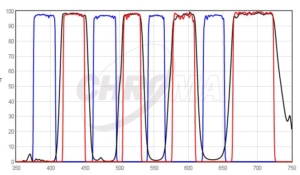
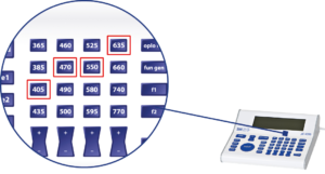
 

DAPI/FITC/TRITC/Cy5 Quad filter excited by pE-4000 at matched wavelengths shown above.

CoolLED’s innovation comes from recognizing that all the stains used in multi-band work can have their absorption bands divided into four separate groups across the spectrum due to the restricted bandwidth available.  This has allowed the development of a patent-pending, wavelength-grouping concept which makes it possible to deliver more power in an efficient four channel system.
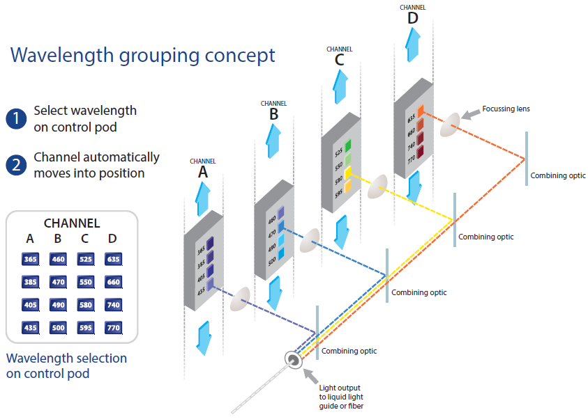
 

With its unique 16 selectable LED wavelengths, the pE-4000 ensures optimum compatibility with all single and multi-band filter sets.  The flexibility and extensive functionality of the pE-4000 broadens the range of illumination options in core facilities.
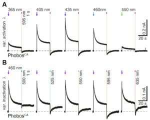
 

The pE-4000 allows multiple experiments with varying wavelengths on the same sample , as shown here.

(A) Representative photocurrent traces of a PhobosCA expressing CA1 cell evoked with different activation wavelengths and shutoff with 595nm light

(B) Photocurrent traces in the same cell evoked with 460nm light and shutoff with indicated wavelengths (10mW/mm2)

Wietek et al (2017) Anion-conducting channelrhodopsins with tuned spectra and modified kinetics engineered for optogenetic manipulation of behaviour,  Scientific Reports volume 7, Article number 14957(2017) doi: 10.1038/s41598-017-14330-y


SUSTAINABILITY

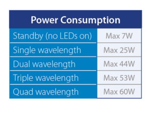
  • Lowest Power Consumption – other LED technology uses 120-350 Watts
  • >25000 hours operation
  • Highly stable, repeatable LED technology
  • Market leading energy efficiency


CELL VIABILITY

  • Optimal illumination through control features to extend fluorescence of cells
  • Reduced photobleaching and phototoxicity
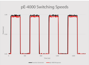
  • Microsecond switching
  • Variable pulse duration on/off

“When you can only control intensity of ‘white’ light (rather than individual channels), the level of photobleaching can be high.  With the pE-4000, we can control the excitation of the individual channels.  It is possible to optimise the excitation intensity according to the labelling, greatly reducing photobleaching and phototoxicity in a live experiment.”    Dr Yan Gu, University of Sussex

“Suddenly, we were able to offer users uninterrupted extended live cell experiments of 100+ hours, without worrying about brightness fluctuations, lamps burning, room heating, etc. Also, users have reported markedly reduced bleaching and phototoxicity in their samples, both from the prokaryotic and the eukaryotic research fields.”     Dr Jens Eriksson, Oslo University Hospital


CONTROLLABILITY

  • Advance integration into major microscope software platforms
  • Save wavelength combinations and return to previous set-up
  • USB interface
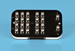
  • High speed TTL Triggering
  • Analogue control – dynamic intensity sinusoidal control option
  • White light control
  • Expansion Box
  • Individual channel intensity control
  • Pre-determined intensity saving option
 

VISIBILITY

  • High signal to noise ratio which gives cleaner images and data

“Striking, bright fluorescence images with a strong signal to noise ratio even when using a low magnification objective” 

Graham Wright, Institute of Medical Biology, A*STAR Singapore
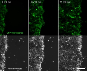
Time series of a confluent sheet of keratinocytes migrating during a live-cell imaging outgrowth assay, acquired on an Olympus IX-83 microscope equipped with a CoolLED pE-4000 lightsource (Fig. 4). Scale bar = 150 μm


ADAPTABILITY

  • Fits to all major research and clinical microscopes
  • Delivers light through a liquid light guide or fiber
  • Offers inline removable excitation filter holders
  • Uses the Universal Collimator


RELIABILITY

  • Assured through experience and QA assured processes
  • 3 year Warranty after registration
  • First year Warranty swap

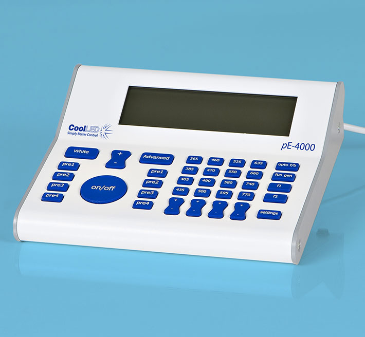
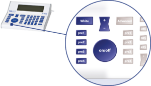
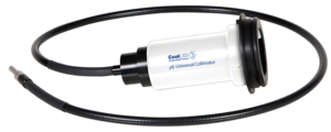
The compact pE-4000 Light Source houses all the LEDs, their combining optics, thermal management and control electronics.  A dual mode control pod gives manual control in either White or Advanced modes.  Light is delivered to the microscope by either a single liquid light guide (LLG) or fiber.  LLGs can be used in conjunction with CoolLED’s pE-Universal Collimator which can accept a microscope adaptor from CoolLED’s extensive range.
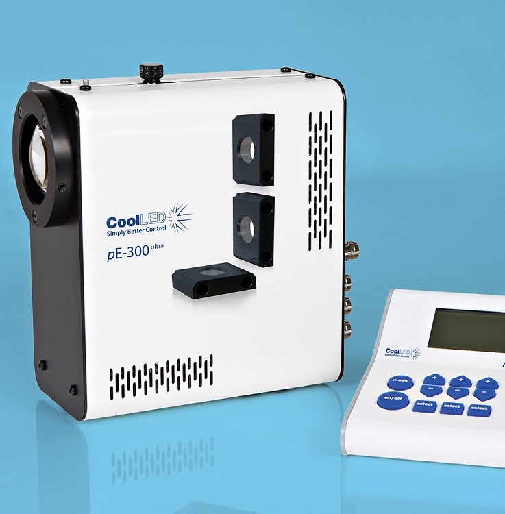
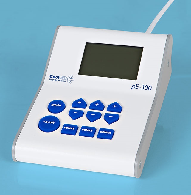


PE-300ultra


CoolLED pE-300ultra


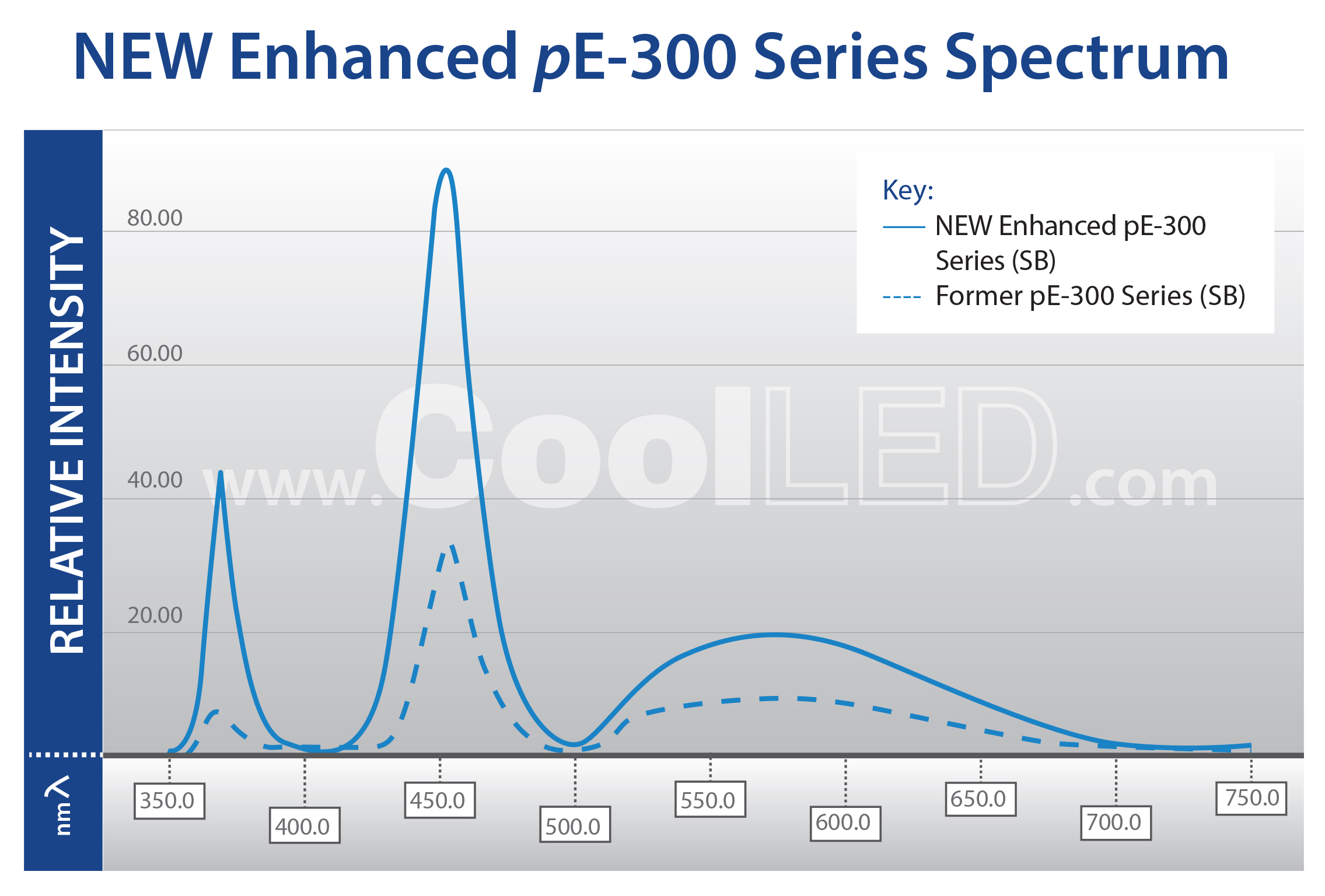
NOW DOUBLE THE INTENSITY AT THE SAMPLE PLANE

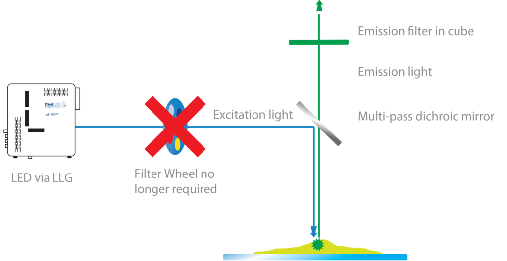
CoolLED’s pE-300ultra is a fluorescence microscopy light source which offers intense, broad-spectrum LED illumination for imaging most common fluorescent stains.  Users have access to both microsecond switching via multiple TTL inputs and the ability to mount inline excitation filters.  This, when paired with today’s high performance multi band filter sets, facilitates imaging traditionally done via a white light source and a filter wheel, with all the benefits of LED.


The pE-300ultra is the most controllable member of the pE-300 Series.  In addition to the many features of the pE-300lite and the pE-300white, the system offers precise control over wavelength intensity and shuttering.  Until now these benefits have only been accessible to users of high end, highly flexible illuminators such as the CoolLED pE-4000.

Throughout 2017 we will be releasing exciting new products and product updates.  The first member of the CoolLED product range to receive the star treatment is the pE-300 Series.

As part of the award winning pE-300 Series, the NEW Enhanced pE-300ultra  now delivers DOUBLE the intensity at the microscope sample plane.  It allows adjustment of output in 1% steps, giving precise control.  The broad spectrum covers everyday fluorophores: DAPI, CFP, Aqua, FITC, TRITC, TxRed, Cy5 and many more.

It is now possible to view and image your samples brighter than ever.

Convaleria taken with Photometrics Prime camera with 10ms exposure.


LED illumination means no mercury to dispose of and lower energy costs due to low power consumption and the precise control offered. With the new enhanced pE-300ultra, all 3 channels at full power use just 46 Watts. That is almost a quadruple efficiency increase!

This compares to other LED technology that uses 120 to 350 Watts. This leap forward in technology makes LEDs more attractive when compared to old Mercury or Metal Halide technology.  Where “green” funding is available, the reduction in institutional energy usage improves the return on investment.

CoolLED’s pE-300 Series  was the 2017 winner of the “Go Beyond Award” in the “Products” category for “Excellence in sustainability in laboratory and other high technology facilities”  The International Institute for Sustainable Laboratories (I2SL) runs a unique awards program honouring organisations, individuals, products,and projects that are advancing sustainable, high-performance facilities.

The pE-300ultra  offers triggering from multiple TTL inputs which, coupled with the ability to mount inline excitation filters, provides microsecond switching of pre-filtered excitation light.  This, when paired with today’s high performance multi-band filter sets, facilitates imaging traditionally done using a white light source and a filter wheel, with all the benefits of LED, and most excitingly at speeds not previously so affordable.

The system comprises a pE-300ultra Light Source, Control Pod, set of three Excitation Filter Holders and Power Supply.  It can be specified with a microscope adaptor for direct fit configuration or with a 3mm diameter liquid light guide if there is a need to keep the source of illumination remote from the microscope.  An optional pE-Universal Collimator and microscope adaptor can be selected for use with a liquid light guide.

A benefit of the pE-300ultra is that  stains can be viewed either individually or in combination, without filter cube changes.  This makes it ideal for use with multi-band filter sets as the screening process can be simplified when fewer filter cubes are used.  Independent control of the three LED channels means that the user can control the level of excitation of each fluorescent stain independently on a multi-stained sample, potentially removing the need for single band filter sets altogether. For further information on improved multi-stain testing, go to the Downloads tab.

An important additional feature of the pE-300ultra is that it includes CoolLED’s “Sequence Runner” multiple channel excitation mode.  Users can define the order of their fluorophore capture using their pE-300ultra Control Pod, then the pE-300ultra light source can accept a single TTL output from the experiment set-up’s camera to initiate the step-through of a sequence of excitation channels.  This feature is independent of the individual channel TTL inputs on the light source.  This offers users the facility to run through a sequence of excitation channels using a camera which has only a single TTL-out.

With an expected lifetime in excess of 25,000 on hours and a comprehensive range of microscope adaptors, the pE-300ultra can be fitted to most current and older microscopes and operate for many years without aligning or replacing bulbs. The result is a safe, convenient illumination system without any additional operating costs.


There are two pE-300ultra configuration options:

Direct-fit for connecting to a microscopes – by selecting from a range of microscope adaptors  which covers all current and most older models.  A simple once only adjustment will allow optimisation to the optical path of the microscope.

Liquid Light Guidewith a fixed 3mm diameter, liquid light guide.  An optional pE-Universal Collimator can be specified in conjunction with a microscope adaptor if required.


The pE-300ultra offers:

  • Clean bright illumination across the spectrum – excites common fluorescent stains
  • Specify for  existing single and multi-band filter sets – no need to buy new filters
  • Instant on/off – No shutters required, no warm up or cool down
  • Simple to fit, simple to use – no alignment, a once only adjustment
  • Stable & repeatable – reliable and consistent results
  • Wide range of microscope adaptors – fits most microscopes
  • Precise intensity control in 1% steps (0-100%) – no ND filters required
  • Excellent uniformity over field of view – fixed and stable, no alignment necessary
  • Long lifetime – expected to exceed 25,000 hours of operating time
  • Removable inline excitation filter holders – no moving parts
  • Individual channel triggering via TTL in microseconds
  • Sequence Runner – sequenced excitation from a single TTL-out


Fluorophores

Browse By Fluorochrome

Compare filter sets by selecting the recommended/alternative set listed for each fluorochrome below.

  • If you need more information, please click fluorochrome. You can see each fluorochrome information and recommended filter sets.
  • If you need other dye information, please contact us.
  • Tel : 02-3473-4188~9
  • E-mail : jhjin@jnoptic.com

하단의 Fluorochrome 이름을 클릭하시면
대응되는 형광필터의 조합을 확인 하실 수 있습니다.

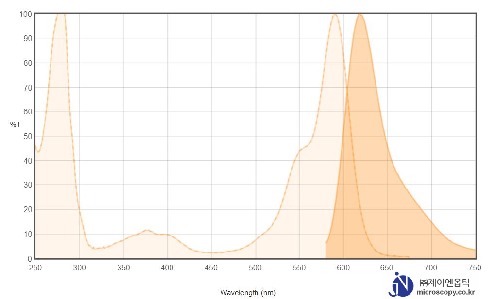
AlexaFluor 594

Alexa Fluor 594 dye is a bright, red-fluorescent dye that can be excited using the 561 nm or 594 nm laser lines. For stable signal generation in imaging and flow cytometry, Alexa Fluor 594 dye is pH-insensitive over a wide molar range.

Spectrum-Chart_Alexa-Fluor-594

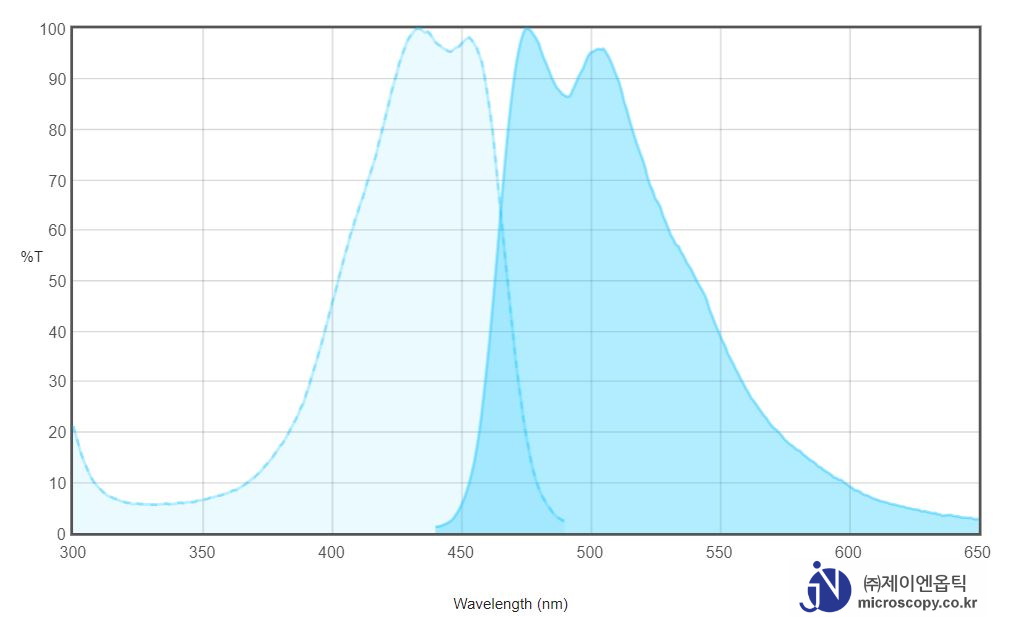
CFP

Cyan Fluorescent Protein (CFP) is a versatile biological marker for monitoring physiological processes, visualizing protein localization, and detecting transgenic expression in vivo. CFP can be excited by the 405 nm laser line and is optimally detected at 485 nm.

Spectrum-Chart_CFP
Spectrum-Chart_CFP

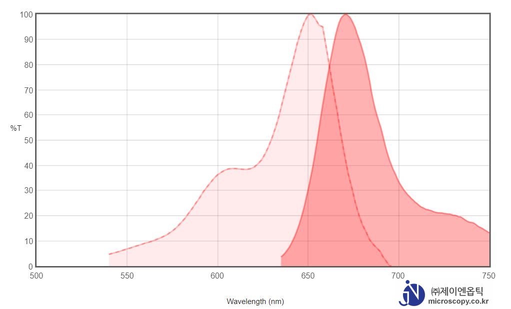
Cy5

Cy5 dye is a bright, far-red-fluorescent dye that can be excited using the 633 or 647 nm laser lines, and visualized Cy5 filter sets.  In addition to immunocytochemistry applications, Cy5 is also commonly used to label nucleic acids.

Spectrum-Chart_Cy5
Spectrum-Chart_Cy5

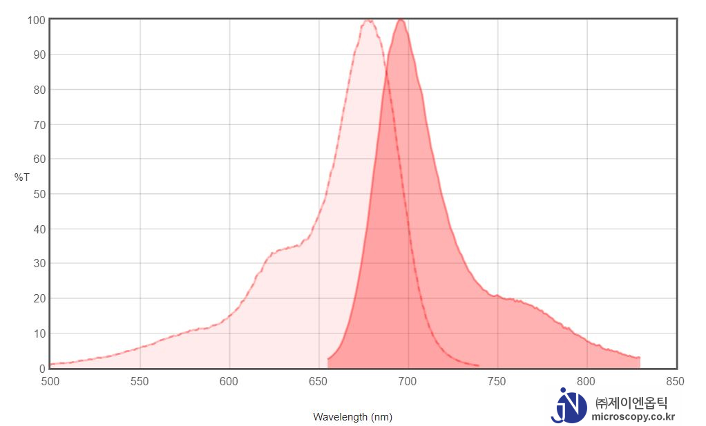
Cy5.5

A variety of cyanine 5.5 (Cy5.5) dyes has been used to label biological molecules for fluorescence imaging and other fluorescence-based biochemical analysis. They are widely used for labeling peptides, proteins and oligos etc. Cy5.5 dyes are one type of the most common red fluorophores.

Spectrum-Chart_Cy5.5

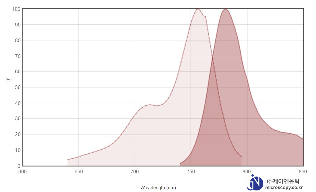
Cy7

Cy7 is a near-IR fluor that is invisible to the naked eye (Excitation/emission maximum 750/776 nm). It is used in in vivo imaging applications, as well as the Cy7.5 dye.

Spectrum-Chart_Cy7
Spectrum-Chart_Cy7

DAPI

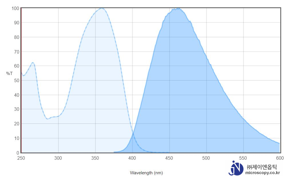
DAPI (4′,6-diamidino-2-phenylindole) is a fluorescent stain that binds strongly to A-T rich regions in DNA. It is used extensively in fluorescence microscopy. As DAPI can pass through an intact cell membrane, it can be used to stain both live and fixed cells, though it passes through the membrane less efficiently in live cells and therefore the effectiveness of the stain is lower.

Spectrum-Chart_DAPI
Spectrum-Chart_DAPI

FITC

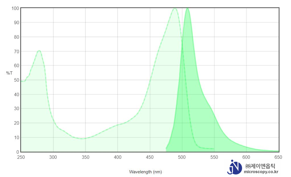
Fluorescein isothiocyanate (FITC) is a derivative of fluorescein used in wide-ranging applications including flow cytometry. FITC is the original fluoresce in molecule functionalized with an isothiocyanate reactive group (-N=C=S), replacing a hydrogen atom on the bottom ring of the structure. This derivative is reactive towards nucleophiles including amine and sulfhydryl groups on proteins.

Spectrum-Chart_FITC
Spectrum-Chart_FITC

GFP

The green fluorescent protein (GFP) is a protein composed of 238 amino acid residues (26.9 kDa) that exhibits bright green fluorescence when exposed to light in the blue to ultraviolet range. Although many other marine organisms have similar green fluorescent proteins, GFP traditionally refers to the protein first isolated from the jellyfish Aequorea victoria. The GFP from A. victoria has a major excitation peak at a wavelength of 395 nm and a minor one at 475 nm. Its emission peak is at 509 nm, which is in the lower green portion of the visible spectrum.

Spectrum-Chart_GFP
Spectrum-Chart_GFP

tdTomato

tdTomato is a genetic fusion of two copies of the dTomato gene (2) which was specifi cally designed for low aggregation (1). Its tandem dimer structure plays an important role in the exceptional brightness of tdTomato (Table I). Its excitation and emission maxima occur at 554 nm and 581 nm, respectively (1). Because tdTomato forms an intramolecular dimer, it behaves like a monomer, and has been used successfully for N- and C- terminal fusions. It shows excellent photostability and its half-time (t0.5) for maturation is one hour at 37°C.

TRITC

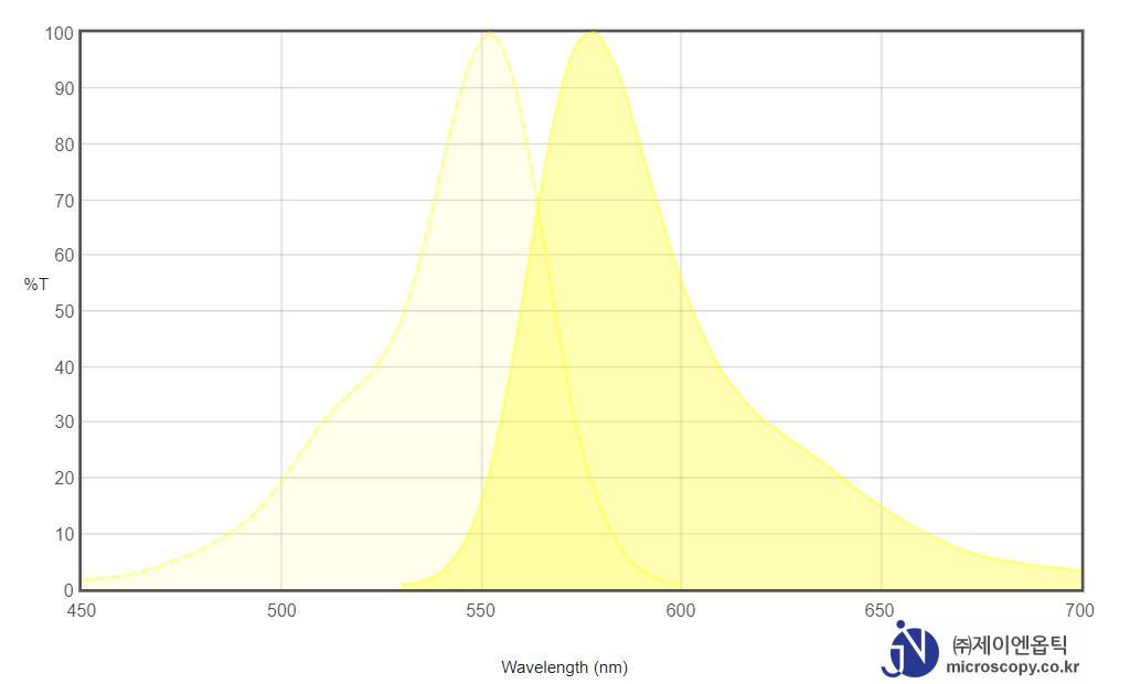
Tetramethylrhodamine (TRITC) is a bright orange-fluorescent dye with excitation ideally suited to the 532 nm laser line. It is commonly conjugated to antibodies and proteins for cellular imaging applications.

Spectrum-Chart_TRITC

light source for Fluorescence

형광 관찰 조명장치의 종류

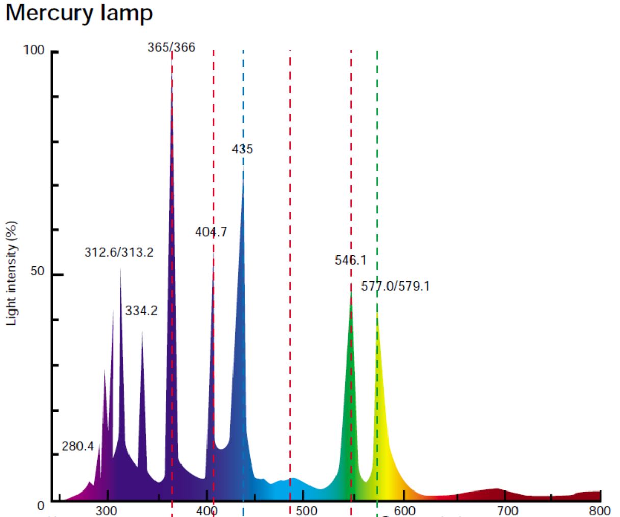
  • Mercury : 형광 현미경법에 가장 흔히 사용하는 광원으로, 자외선 및 가시광선 영역에서 아주 밝다.
Spectrum of mercury
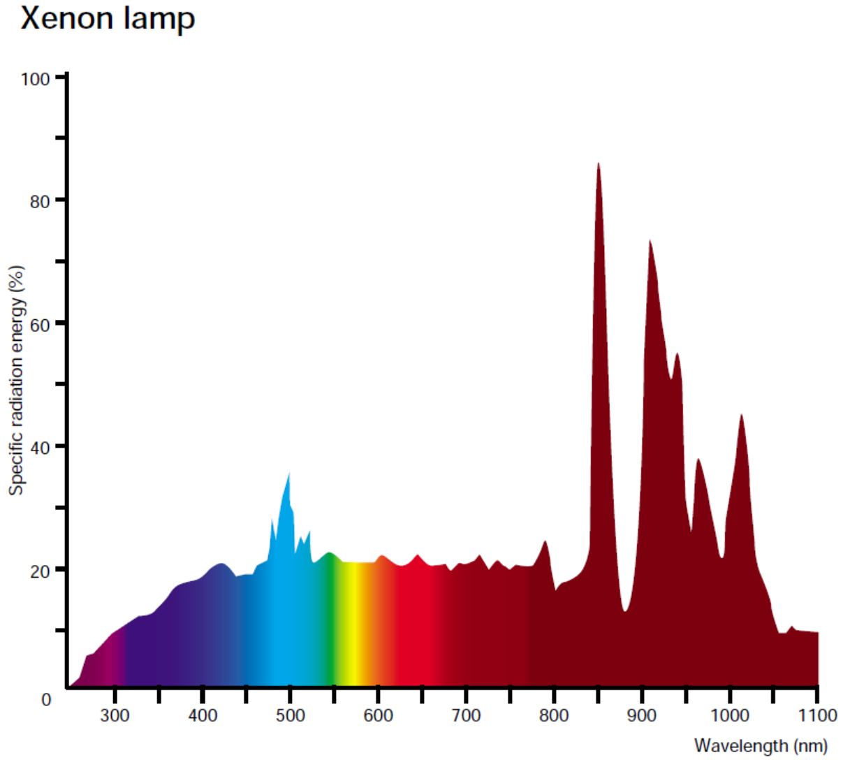
  • Xenon : 가시광선에서 상대적으로 연속적인 스펙트럼을 가진다. 염료 및/또는 시료의 스펙트럼 특성을 정량적으로 분석하는 시스템에서는 제논 램프를 사용하는 것이 좋지만, 같은 와트수의 수은 램프만큼 밝지는 않다.
Spectrum of Xenon
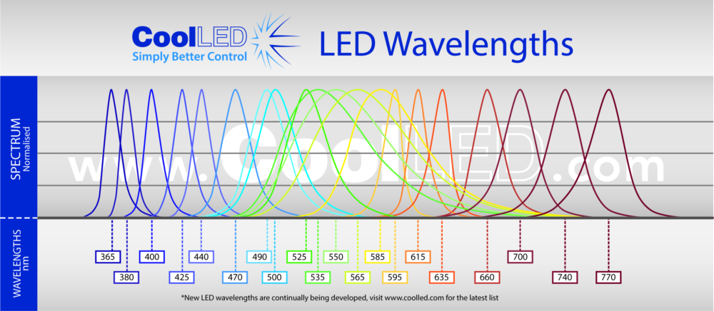
  • LED :  약 10 ~ 40nm 범위의 상대적으로 좁은 스펙트럼 대역폭 내에서 빛을 방출한다는 고유의 특징이 있다. 일반적으로 정격 수명이 10,000시간 이상이고 50,000시간 이상 사용하는 경우도 많고, 전체 정격 수명에 걸쳐 출력 강도가 비교적 일정하게 유지된다.
Spectrum of LED

Laser : 레이저

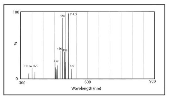
  • 보통은 단색광의 좁은 스펙트럼 대역에서 극히 밝다. 하지만 레이저광의 간섭성으로 인해 샘플 영역에 균일한 조도로 비추기 어렵지만, Confocal 현미경에서는 갈바노 미러를 이용한 스캔 방식을 이용하여 이미지 획득시에 사용되어진다(LSCM).
    Spectrum of Laser

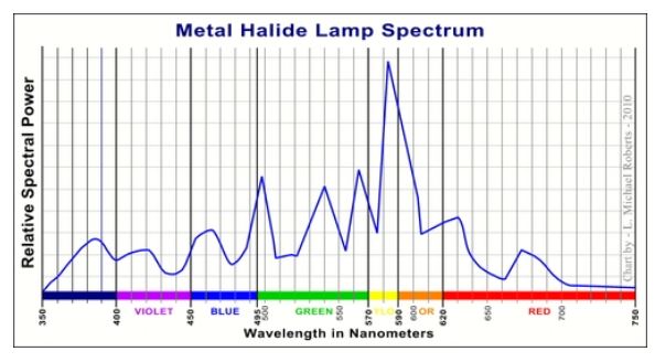
Metal-halide : 

  • 유효수명이 최대 2000시간으로, 일반적인 수은 램프의 200시간이나 제논 램프의 400시간과 비교하면 훨씬 길다.

JNO-G(L)

JNOPTIC Filter JNO-G(L)

– Filter set for TRITC / Cy3 Longpass
– Chroma Filter Model No. : 19004

JNO-G(L) Spectrum

Standard Green Excitaion filter (Long Pass type)
FiltersType
AT540/25xEX
AT565DCBS
AT575lpEM

주요 형광 색소(Fluorochromes)와 스펙트럼 비교

* TRITC VS JNO-G(L)

TRITC VS JNO-G(L)
TRITC VS JNO-G(L)

JNO-G(L)필터를 이용한 Sample Image 촬영

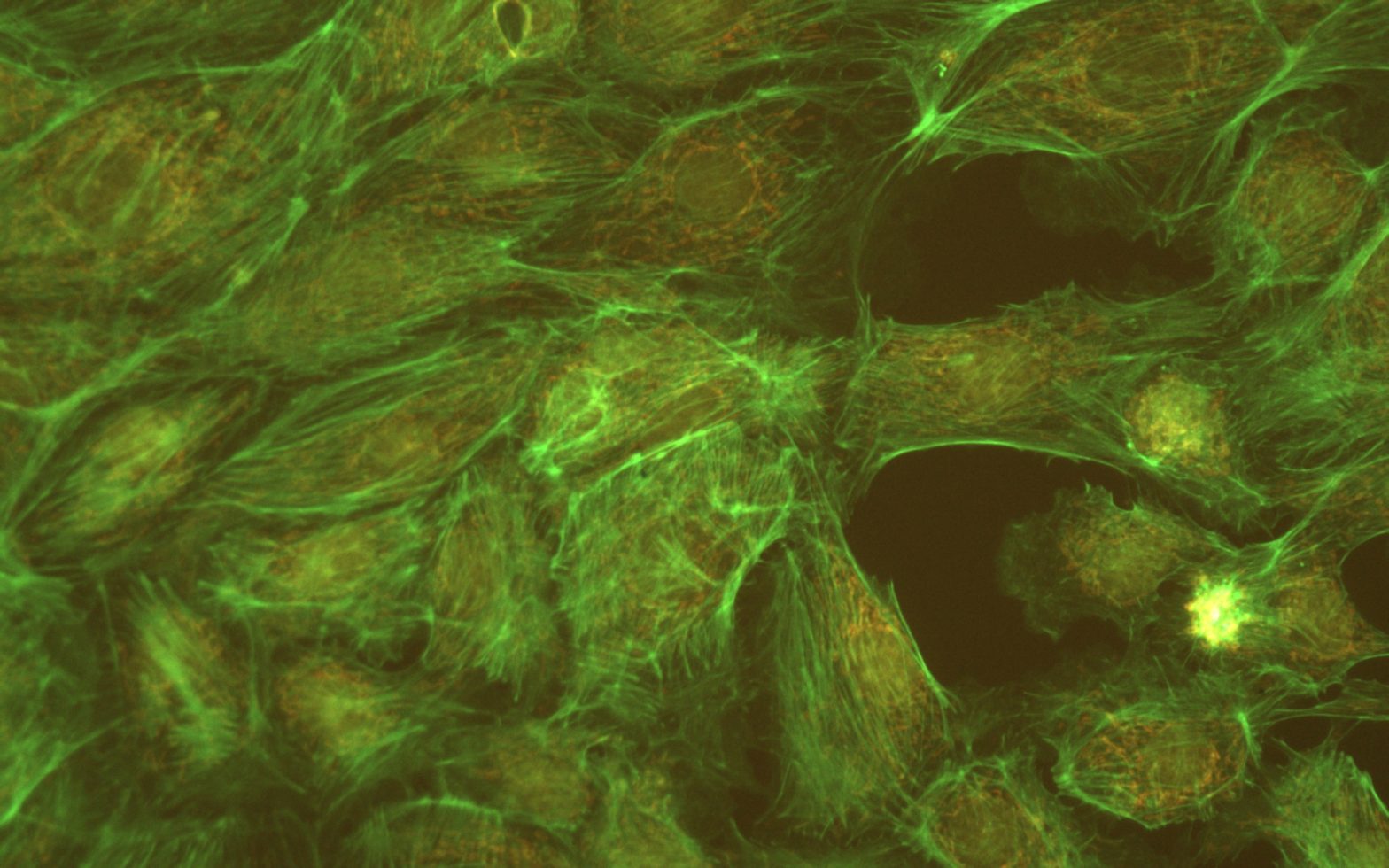
Fluorescence image taken using JNO-G(L)
Filter Set : JNO-G(L) Camera : AcquCAM 23GR2 C-mount adapter : 1x Lens : UPLAPO40X Sample : bovine pulmonary artery endothelial cell, BPAEC
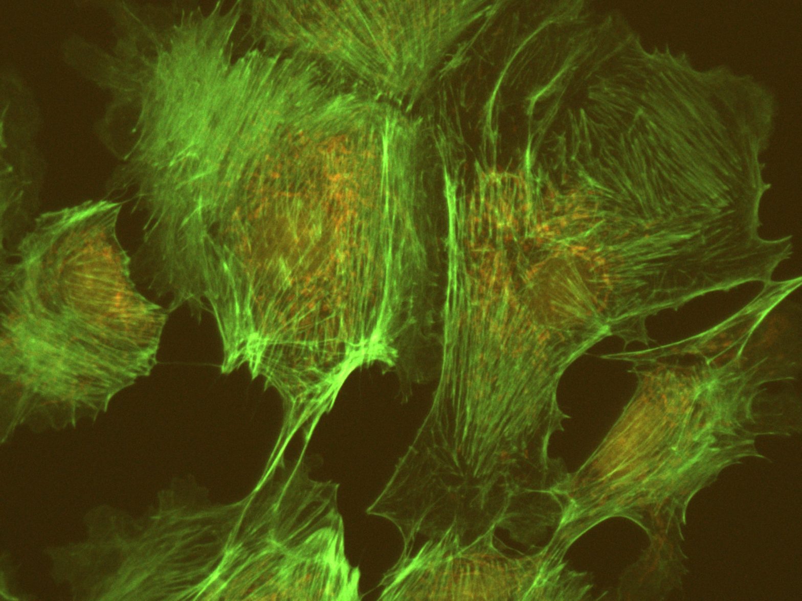
Fluorescence image taken using JNO-G(L)
Filter Set : JNO-G(L) Camera : AcquCAM 5G C-mount adapter : 1x Lens : UPLAPO40X Sample : bovine pulmonary artery endothelial cell, BPAEC
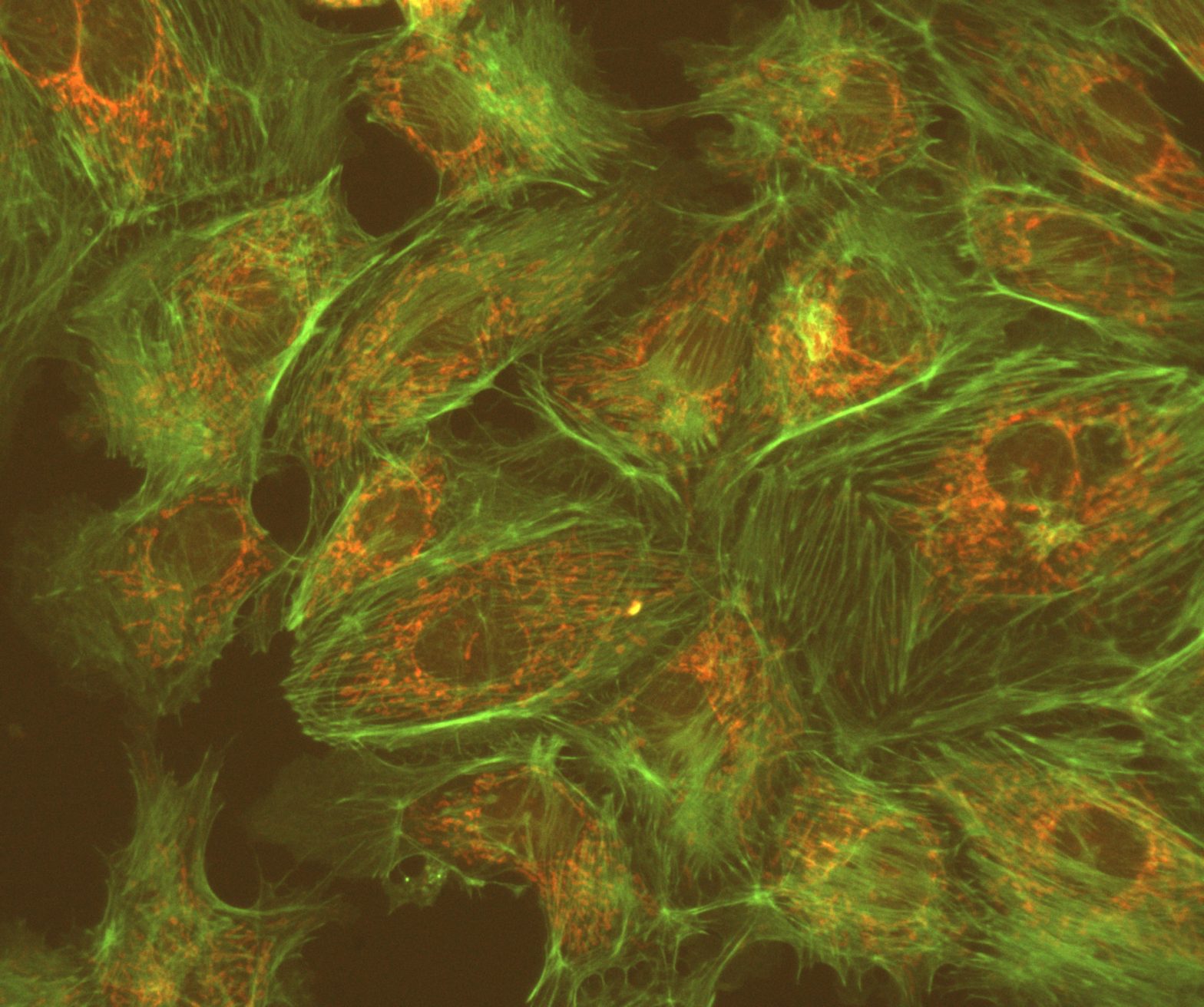
Fluorescence image taken using JNO-G(L)
Filter Set : JNO-G(L) Camera : AcquCAM 3G C-mount adapter : 1x Lens : UPLAPO40X Sample : bovine pulmonary artery endothelial cell, BPAEC

JNO-G(L)필터에 적합한 형광색소

  • R-phycoerythrin

JNO-G(L) Spec

  • Typical Application(s) : Widefield Microscopy
  • Coating : Sputter/Hard Coated
  • Round excitation and emission filters mounted in anodized aluminum rings 2.3mm thick, up to 25mm in diameter (for standard sizes)
  • Rectangular dichroics up to 26x38mm, 1mm thick, to fit standard microscope manufacturer filter cubes (for standard sizes)

*** For filters larger than 25mm and dichroics larger than 26x38mm, please contact us for pricing.

JNO-B(L)

JNOPTIC Filter JNO-B(L)

– Filter set for GFP / FITC Longpass
– Chroma Filter Model No. : 19002

JNO-B(L) Spectrum

JNO-B(L) Spectrum
JNO-B(L) Spectrum
FiltersType
AT480/30xEX
 AT505DCBS
AT515lpEM

주요 형광 색소(Fluorochromes)와 스펙트럼 비교

 * FITC VS JNO-B(L)

FITC VS JNO-B(L)
FITC VS JNO-B(L)

 * GFP VS JNO-B(L)

GFP VS JNO-B(L)
GFP VS JNO-B(L)

JNO-B(L)필터를 이용한 Sample Image 촬영

Fluorescence image taken using JNO-B(L)
Sample : bovine pulmonary artery endothelial cell, BPAEC Filter Set : JNO-B(L) Camera : AcquCAM 23GR2 C-mount adapter : 1X Lens : UPLNAPO40X
Fluorescence image taken using JNO-B(L)
Sample : bovine pulmonary artery endothelial cell, BPAEC Filter Set : JNO-B(L) Camera : AcquCAM 3G C-mount adapter : 1X Lens : UPLNAPO40X
Fluorescence image taken using JNO-B(L)
Sample : bovine pulmonary artery endothelial cell, BPAEC Filter Set : JNO-B(L) Camera : AcquCAM 5G C-mount adapter : 1X Lens : UPLNAPO40X

JNO-B(L)필터에 적합한 형광색소

  • Acridine Orange + DNA and RNA
  • Auramine-rhodamine
  • JC-1 non-ratiometric

JNO-B(L) Spec

  • Typical Application(s) : Widefield Microscopy
  • Coating : Sputter/Hard Coated
  • Round excitation and emission filters mounted in anodized aluminum rings 2.3mm thick, up to 25mm in diameter (for standard sizes)
  • Rectangular dichroics up to 26x38mm, 1mm thick, to fit standard microscope manufacturer filter cubes (for standard sizes)

*** For filters larger than 25mm and dichroics larger than 26x38mm, please contact us for pricing. 02-3473-4188~9

JNO-U(L)

JNOPTIC Filter  JNO-U(L)

– Filter set for UV/DAPI Longpass
– Chroma Filter Model No. : 19000

JNO-U(L) Spectrum

JNO-U(L) Spectrum Chart
FiltersType
AT375/28xEX
AT415DCBS
AT435lpEM

주요 형광 색소(Fluorochromes)와 스펙트럼 비교

* DAPI VS JNO-U(L)

DAPI VS JNO-U(L)
DAPI VS JNO-U(L)

JNO-U(L)필터를 이용한 Sample Image 촬영

Fluorescence image taken using JNO-U(L)
DAPI Sample : bovine pulmonary artery endothelial cell, BPAEC Lens : UPLNAPO40X, C-mount adapter : 1X Camera : AcquCAM 23GR2, Filter Set : JNO-U(L)
Fluorescence image taken using JNO-U(L)
DAPI Sample : bovine pulmonary artery endothelial cell, BPAEC Lens : UPLNAPO40X, C-mount adapter : 1X Camera : AcquCAM 3G, Filter Set : JNO-U(L)
Fluorescence image taken using JNO-U(L)
DAPI Sample : bovine pulmonary artery endothelial cell, BPAEC Lens : UPLNAPO40X, C-mount adapter : 1X Camera : AcquCAM 5G, Filter Set : JNO-U(L)

JNO-U(L)에 적합한 형광색소

  • Calcofluor White
  • DAPI

JNO-U(L) Spec

  • Typical Application(s) : Widefield Microscopy
  • Coating : Sputter/Hard Coated
  • Round excitation and emission filters mounted in anodized aluminum rings 2.3mm thick, up to 25mm in diameter (for standard sizes)
  • Rectangular dichroics up to 26x38mm, 1mm thick, to fit standard microscope manufacturer filter cubes (for standard sizes)

*** For filters larger than 25mm and dichroics larger than 26x38mm, please contact us for pricing.

JNO-R2(B)

JNOPTIC Filter  JNO-R2(B)

– Filter set for Cy5 / AlexaFluor 647 / Draq5
– Chroma Filter Model No. : 39007

JNO-R2(B) Spectrum

FiltersType
 AT620/50xEX
AT655DCBS
AT690/50mEM

주요 형광 색소(Fluorochromes)와 스펙트럼 비교

* Cy5 VS JNO-R2(B)

Cy5 VS JNO-R2(B)
Cy5 VS JNO-R2(B)

 * AlexaFluor 647 VS JNO-R2(B)

AlexaFluor 647 VS JNO-R2(B)

 * Draq5 VS JNO-R2 (B)

Draq5 VS JNO-R2 (B)
Draq5 VS JNO-R2 (B)

JNO-R2(B)필터에 적합한 형광색소

  • Alexa Fluor 647™
  • Atto 647N
  • Cy5™
  • DiD
  • Draq5
  • DyLight 649
  • MitoTracker Deep Red 633/MeOH
  • Nile Blue
  • Quasar® 670
  • SYTO® 60
  • TO-PRO™-3

JNO-R2(B) Spec

  • Typical Application(s) : Widefield Microscopy
  • Coating : Sputter/Hard Coated
  • Round excitation and emission filters mounted in anodized aluminum rings 2.3mm thick, up to 25mm in diameter (for standard sizes)
  • Rectangular dichroics up to 26x38mm, 1mm thick, to fit standard microscope manufacturer filter cubes (for standard sizes)

*** For filters larger than 25mm and dichroics larger than 26x38mm, please contact us for pricing.

JNO-R1(B)

JNOPTIC Filter JNO-R1(B)

– Filter set for Texas Red / mCherry / AlexaFluor 594
– Chroma Filter Model No. : 39010

JNO-R1(B) Spectrum

JNO-R1(B) Spectrum
JNO-R1(B) Spectrum
FiltersType
AT560/40xEX
AT600DCBS
AT635/60mEM

주요 형광 색소(Fluorochromes)와 스펙트럼 비교

* AlexaFluor 594 VS JNO-R1(B)

Alexa Fluor 594 VS JNO-R1(B)

 * Texas Red VS JNO-R1(B)

Texas Red VS JNO-R1(B)

 * mCherry VS JNO-R1(B)

mCherry VS JNO-R1(B)

JNO-R1(B)필터에 적합한 형광색소

  • Alexa Fluor 568™
  • Alexa Fluor 594™
  • CAL Fluor® Red 610
  • Cy3.5™
  • DyLight 594
  • FusionRed
  • Killer Red
  • mCherry
  • MitoTracker Red/MeOH
  • mKate2
  • mRFP1
  • ReAsH-CCPGCC
  • Rhodamine Red™-X
  • ROX
  • Sulforhodamine 101
  • Texas Red®
  • Texas Red®-X
  • X-rhod-1/Ca2+

JNO-R1(B) Spec

  • Typical Application(s) : Widefield Microscopy
  • Coating : Sputter/Hard Coated
  • Round excitation and emission filters mounted in anodized aluminum rings 2.3mm thick, up to 25mm in diameter (for standard sizes)
  • Rectangular dichroics up to 26x38mm, 1mm thick, to fit standard microscope manufacturer filter cubes (for standard sizes)

*** For filters larger than 25mm and dichroics larger than 26x38mm, please contact us for pricing.

JNO-G(B)

JNOPTIC Filter JNO-G(B)

– Filter set for TRITC / Cy3 / TagRFP / AlexaFluor546
– Chroma Filter Model No. : 39004

JNO-G(B) Spectrum

JNO-B(L) Spectrum
JNO-B(L) Spectrum
FiltersType
AT540/25xEX
AT565DCBS
AT605/55mEM

주요 형광 색소(Fluorochromes)와 스펙트럼 비교

* TRITC VS JNO-G(B)

TRITC에 사용했을때 JNO-G(B)

 * Cy3 VS JNO-G(B)

Cy3에 사용되는 JNO-G(B)의 스펙트럼 비교

 * AlexaFluor546 VS JNO-G(B)

AF594에 사용되는 JNO-G(B)의 스펙트럼 비교

JNO-G(B)필터를 이용한 Sample Image 촬영

Fluorescence image taken using JNO-G(B)
Filter Set : JNO-G(B) Camera : AcquCAM 3G C-mount adapter : 1x Lens : UPLAPO20X Sample : bovine pulmonary artery endothelial cell, BPAEC
Fluorescence image taken using JNO-G(B)
Filter Set : JNO-G(B) Camera : AcquCAM 5G C-mount adapter : 1x Lens : UPLAPO40X Sample : bovine pulmonary artery endothelial cell, BPAEC
Fluorescence image taken using JNO-G(B)
Filter Set : JNO-G(B) Camera : AcquCAM 23GR2 C-mount adapter : 1x Lens : UPLAPO20X Sample : bovine pulmonary artery endothelial cell, BPAEC

JNO-G(B)필터에 적합한 형광색소

  • Alexa Fluor 546™
  • Alexa Fluor 555™
  • Atto 550
  • CAL Fluor® Red 590
  • Cy3™
  • DiI
  • DsRed
  • DyLight 549
  • MitoTracker Orange/MeOH
  • Resorufin
  • Rhod-2
  • TagRFP
  • TAMRA
  • tdTomato
  • Tetramethylrhodamine isothiocyanate
  • TRITC

JNO-G(B) Spec

  • Typical Application(s) : Widefield Microscopy
  • Coating : Sputter/Hard Coated
  • Round excitation and emission filters mounted in anodized aluminum rings 2.3mm thick, up to 25mm in diameter (for standard sizes)
  • Rectangular dichroics up to 26x38mm, 1mm thick, to fit standard microscope manufacturer filter cubes (for standard sizes)

*** For filters larger than 25mm and dichroics larger than 26x38mm, please contact us for pricing.